A Rash of Beta-Lactam Allergies, Part 2: The Education

This post is the second in a three-part series covering the management of beta-lactam allergies, all to be released on FOAMid over the last few months of 2019. Part 1 explained the enormous impact that penicillin allergies have on patient outcomes. Today we’ll discuss the different types of allergic reactions and the potential for beta-lactam allergy cross reactivity. Let’s jump right in!

Types of Allergic Reactions

The most common way of grouping immune-mediated hypersensitivity reactions is through the Gell & Coombs classification method.2 Using this scheme, there are four types of allergic reaction:

Type I reactions are IgE-mediated reactions and commonly referred to as immediate-type hypersensitivity reactions, since they occur minutes to hours post-exposure to an allergen. Type I reactions include anaphylaxis, angioedema, hypotension, flushing, wheezing, hives, and urticaria.

Both types II and III reactions are IgG-mediated.
Type II reactions, or cytotoxic reactions, include hemolytic anemia, thrombocytopenia, and neutropenia.
Type III reactions are immune complex reactions, and include serum sickness, glomerulonephritis, and arthritis.

Last, but certainly not least, are type IV reactions, which are T-cell mediated.
Type IV reactions are commonly referred to as delayed hypersensitivity reactions, despite Types II, III, and IV all technically being delayed in nature by days to weeks post-exposure to an allergen. A maculopapular rash, interstitial nephritis, Stevens-Johnson Syndrome (SJS), toxic epidermal necrolysis (TEN), and drug reaction with eosinophilia and systemic symptoms (DRESS) syndrome are all considered type IV reactions.

Cross-Reactivity Risk

As discussed in the first post, many recorded antibiotic allergies are not true allergies. But when a patient does actually have a true penicillin allergy, what are the chances that the patient will have a similar reaction to other beta-lactams?

While we would generally avoid penicillins in this situation, other beta-lactams like cephalosporins and carbapenems could potentially be used. Previous studies of 10-25% cross-reactivity between cephalosporins and penicillins were primarily reported prior to 1982, when cephalosporin manufacturing processes were often contaminated with penicillin.12 Since then, the documented rate of cross-reactivity has dropped dramatically, shown in the table below.12

*not all cephalosporins are created equal (see comments below)

Cephalosporins

  • Cephalosporins that do not share a side chain with penicillin have a cross-reactivity risk of <2%
  • Cephalosporins that do share a similar side chain to penicillins (ex. cefoxitin and penicillin) have a cross-reactivity risk that is much higher

Side chain similarities don’t guarantee cross-reactivity, but they do increase the risk above the previously stated 2% threshold

But hold on, I thought the cause of beta-lactam allergies was the core beta-lactam ring that everyone remembers from their undergraduate years?

Penicillin molecule with the highlighted beta-lactam ring

Not so fast. While this plays a part, more recent literature has shown that the R1 and R2 side chains also play a role in the allergy potential of cephalosporins. I have adopted and updated a table from an excellent 2008 review paper by Daryl DePestel and colleagues below.3

In this table, the 3s, 6s, and 7s stand for similar R1 or R2 side chains, as described in the cephalosporin skeleton molecule, also seen below.

  • The R1 side chain is at the 7-position on the cephalosporin molecule and the 6-position on the penicillin molecule.
  • The R2 side chain is at the 3-position, which only differs among cephalosporins and not penicillins.

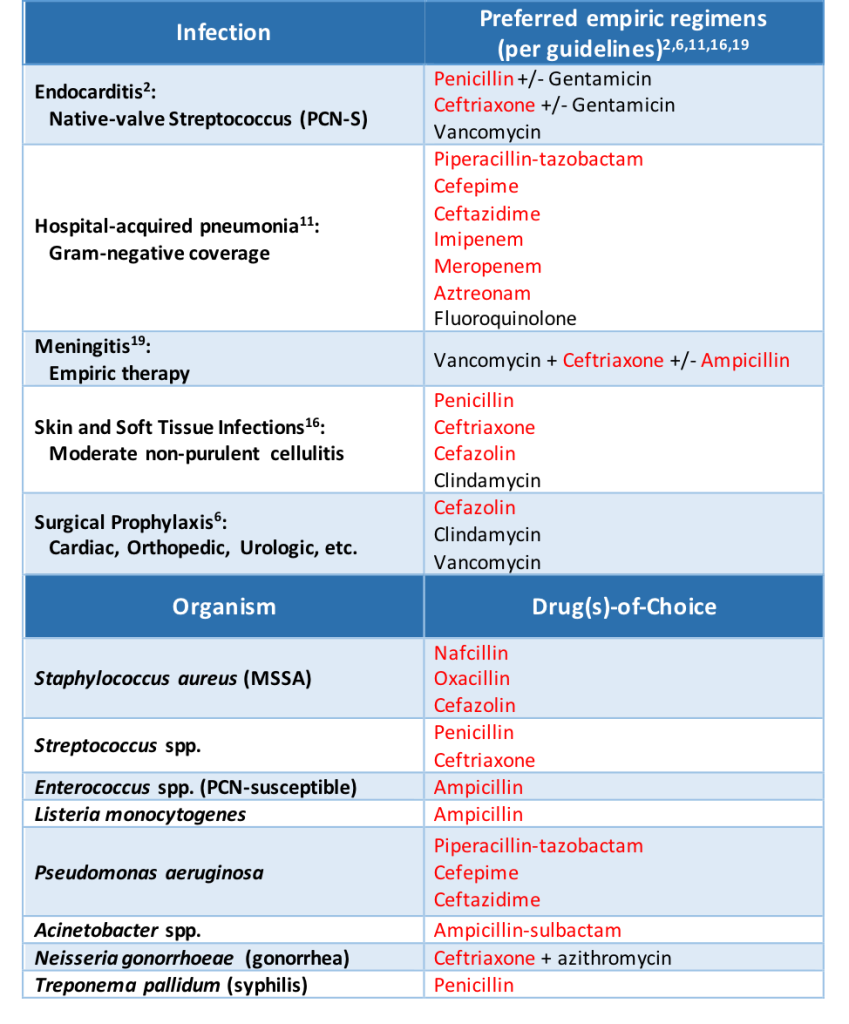
There are a couple of important clinical points to note from this table. Probably most important for clinical practice is that cefazolin does not share side chains with any other beta-lactam agents. This can have huge consequences on the use of cefazolin in practice, especially when it comes to surgical site prophylaxis and the treatment of methicillin-susceptible Staphylococcus aureus infections, both situations that could use cefazolin as first line therapy.

And while aztreonam is known as a beta-lactam with limited cross-reactivity due to dissimilar side chains, it does actually share a side chain with ceftazidime and the more recently approved ceftolozane (marketed in combination with tazobactam).

Aztreonam & Carbapenems

Speaking of aztreonam, we’ve spent the majority of this post discussing cephalosporin cross-reactivity risk. Now let’s spend a bit of time reviewing the other agents defined in the initial table in this post: carbapenems and aztreonam. Cross-reactivity between these agents and penicillins is minimal, as seen by a number of studies published by an Italian group headed by Antonino Romano and Francesco Gaeta.4,7,9

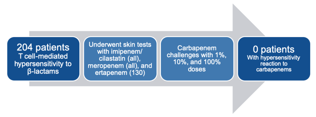
In their 2013 analysis, they found no patients had an allergic reaction to carbapenems, despite all 204 patients having a well-demonstrated T-cell-mediated hypersensitivity reaction to other beta-lactams (mostly penicillin).7

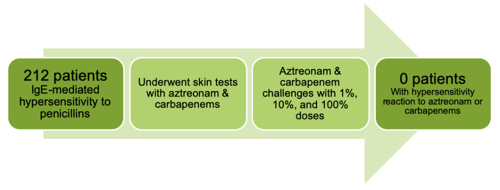
They went on to look at IgE-mediated hypersensitivity in their 2015 study, which found yet again no cases of hypersensitivity with either carbapenems OR aztreonam this time in a cohort of 212 patients with proven penicillin allergies.4

Then in 2016, they went back to T-cell-mediated hypersensitivity, examining 214 patients with proven reactions to penicillins and testing them against aztreonam. Once again, zero patients reacted to the aztreonam test doses or full dose.9

At this point, you may be questioning if the Italian group ever saw any reactions in their trial outcomes. The last study presented above that showed no reactions with aztreonam though tested more than just aztreonam. They also looked at cephalosporins and saw an 18.7% chance of positive skin testing with aminocephalosporins (cephalexin, cefadroxil, cefaclor).9 If you refer back to the previous cross-reactivity table, you can see that these three agents share a side chain with ampicillin and amoxicillin.

So while side chains play a key role in determining cross-reactivity among cephalosporins, we can be fairly confident that carbapenems and aztreonam are safe to administer in the majority of situations, especially when a non-severe penicillin allergy is documented. This will be covered in more detail in the next (and final) installment of “A Rash of Beta-Lactam Allergies,” coming to you soon!

Other posts in this series:

A Rash of Beta-Lactam Allergies, Part 1: The Problem

A Rash of Beta-Lactam Allergies, Part 3: The Solution

References

  1. Blumenthal KG, Peter JG, Trubiano JA, Phillips EJ. Antibiotic Allergy. Lancet. 2019; 393(10167):183-198
  2. Coombs P, Gell PG. Classification of allergic reactions responsible for clinical hypersensitivity and disease. In: G RR, P.G.H Gell, eds. Clinical aspects of immunology. Oxford, UK: Oxford University Press, 1968; 575-596
  3. Frumin J, Gallagher JC. Allergic cross-sensitivity between penicillin, carbapenem, and monobactam antibiotics: what are the chances? Ann Pharmacother. 2009; 43:304-315
  4. Gaeta F, Valluzzi RL, Alonzi C, Maggioletti M, Caruso C, Romano A. Tolerability of aztreonam and carbapenems in patients with IgE-mediated hypersensitivity to penicillins. J Allergy Clin Immunol. 2015; 135:972-976
  5. Joint Task Force on Practice Parameters; American Academy, American College, & Joint Council of Allergy, Asthma and Immunology. Drug allergy: an updated practice parameter. Ann Allergy Asthma Immunol. 2010; 105:259-273
  6. Legendre DP, Muzny CA, Marshall GD, Swiatlo E. Antibiotic hypersensitivity reactions and approaches to desensitization. Clin Infect Dis. 2014; 58(8):1140-1148
  7. Romano A, Gaeta F, Valluzzi RL, et al. Absence of cross-reactivity to carbapenems in patients with delayed hypersensitivity to penicillins. Allergy. 2013; 68:1618-1621
  8. Romano A, Gaeta F, Arribas Poves MF, Valluzzi RL. Cross-reactivity among beta-lactams. Curr Allergy Asthma Rep. 2016; 16:24
  9. Romano A, Gaeta F, Valluzzi RL, Maggioletti M, Caruso C, Quaratino D. Cross-reactivity and tolerability of aztreonam and cephalosporins in subjects with a T cell-mediated hypersensitivity to penicillins. J Allergy Clin Immunol. 2016; 138:179-186
  10. Romano A, Valluzzi RL, Caruso C, Maggioletti M, Quaratino D, Gaeta F. Cross-reactivity and tolerability of cephalosporins in patients with IgE-mediated hypersensitivity to penicillins. J Allergy Clin Immunol Pract. 2018; 6(5):1662-1672
  11. Shenoy ES, Macy E, Rowe T, Blumenthal KG. Evaluation and management of penicillin allergy: a review. JAMA. 2019; 321(2):188-199
  12. Trubiano JA, Stone CA, Grayson ML, et al. The 3 Cs of antibiotic allergy-classification, cross-reactivity, and collaboration. J Allergy Clin Immunol Pract. 2017; 5(6):1532-1542

A Rash of Beta-Lactam Allergies, Part 1: The Problem

This post marks part 1 of a 3-part series covering the management of beta-lactam allergies, all to be released on FOAMid over the next couple of months.

  1.  This post, “The Problem,” provides background and the impact of a reported beta-lactam allergy
  2. “The Education” will delve into the types of allergic reactions, as well as cross reactivity potential among beta-lactam antibiotics
  3. “The Solution” will then explore how to best assess a patient’s documented allergy

With that, let’s jump right in!

Overview

A whopping 10% of the general population has a reported penicillin (PCN) allergy. But only 1-10% of these people have a true allergy when tested. This leaves us with about 0.1-1% of the general population with a true penicillin allergy.

Why is there such a discrepancy between reported allergies and true allergies? A lot of it comes from inaccurate allergy histories, like the patient with GI upset as a child, but the allergy listed as an “unknown reaction.” Or better yet, the patient whose mother had an allergy and thus everyone in the family has been given that scarlet letter in their medical record.

Another important and lesser known reason for the allergy discrepancy is that 78% of patients with immediate hypersensitivity to penicillin see their penicillin allergy fade after 10 years (from this 1981 study). So those adult patients with childhood reactions? The odds are that they aren’t still allergic decades later.

Why should we care?

When it comes to infectious diseases, beta-lactam antibiotics are often our first- and second-line options for treatment. A documented penicillin allergy can essentially knock a practitioner down to third-line treatment in some situations. In just highlighting a few common infections and organisms, look at how often beta-lactams are brought up:

When a patient has a documented penicillin allergy, studies have proven that beta-lactam usage decreases while non-beta-lactam usage increases (Lee 2000, as well as half of the citations provided at the end of this post). And when beta-lactams are avoided, patients tend to do worse.

Impact on Patient Outcomes

The impact of a penicillin allergy is real and detrimental to our patients. Rather than bore you with paragraphs upon paragraphs detailing the many studies looking into this fact, here are some take-home points hyperlinked to the primary literature supporting the claims:

Penicillin allergy patients:

There is clear evidence that reported beta-lactam allergies pose a problem on the path to prescribing optimal treatment in infectious diseases. We can combat the issue however through education and assessment techniques.

More to come in parts 2 and 3 of “A Rash of Beta-Lactam Allergies”!

Other posts in this series:

A Rash of Beta-Lactam Allergies, Part 2: The Education

A Rash of Beta-Lactam Allergies, Part 3: The Solution

References

  1. Al-Hasan MN, Acker EC, Kohn JE, Bookstaver PB, Justo JA. Impact of penicillin allergy on empirical carbapenem use in gram-negative bloodstream infections: an antimicrobial stewardship opportunity. Pharmacotherapy. 2017; 38(1):42-50
  2. Baddour LM, Wilson WR, Bayer AS, et al. Infective endocarditis in adults: diagnosis, antimicrobial therapy, and management of complications: a scientific statement for healthcare professionals from the American Heart Association. Circulation. 2015; 132:1435-1486
  3. Blumenthal KG, Lu N, Zhang Y, Li Y, Walensky RP, Choi HK. Risk of meticillin resistant Staphylococcus aureus and Clostridium difficile in patients with a documented penicillin allergy: population based matched cohort study. BMJ. 2018; 361:k2400
  4. Blumenthal KG, Ryan EE, Li Y, Lee H, Kuhlen JL, Shenoy ES. The impact of a reported penicillin allergy on surgical site infection risk. Clin Infect Dis. 2018; 66(3):329-336
  5. Borch JE, Andersen KE, Bindslev-Jensen C. The prevalence of suspected and challenge-verified penicillin allergy in a university hospital population. Basic Clin Pharmacol Toxicol. 2006; 98:357-362
  6. Bratzler DW, Dellinger EP, Olsen KM, et al. Clinical practice guidelines for antimicrobial prophylaxis in surgery. Surg Infect. 2013;14(1):73-156
  7. Charneski L, Deshpande G, Smith SW. Impact of an antimicrobial allergy label in the medical record on clinical outcomes in hospitalized patients. Pharmacotherapy. 2011; 31(8):742-747
  8. Conway EL, Lin K, Sellick JA, et al. Impact of penicillin allergy on time to first dose of antimicrobial therapy and clinical outcomes. Clin Ther. 2017; 39(11):2276-2283
  9. Huang KHG, Cluzet V, Hamilton K, Fadugba O. The impact of reported beta-lactam allergy in hospitalized patients with hematologic malignancies requiring antibiotics. Clin Infect Dis. 2018; 67(1):27-33
  10. Jeffres MN, Narayanan PP, Shuster JE, Schramm GE. Consequences of avoiding β-lactams in patients with β-lactam allergies. J Allergy Clin Immunol. 2016; 137(4):1148-1153
  11. Kalil AC, Metersky ML, Klompas M, et al. Management of adults with hospital-acquired and ventilator-associated pneumonia: 2016 clinical practice guidelines by the Infectious Diseases Society of America and the American Thoracic Society. Clin Infect Dis. 2016; 63(5):e61-e111
  12. Lee CE, Zembower TR, Fotis MA, et al. The incidence of antimicrobial allergies in hospitalized patients: implications regarding prescribing patterns and emerging bacterial resistance. Arch Intern Med. 2000;160(18):2819-2822
  13. Macy E, Ngor EW. Safely diagnosing clinically significant penicillin allergy using only penicilloyl-poly-lysine, penicillin, and oral amoxicillin. J Allergy Clin Immunol Pract. 2013; 1:258-263
  14. Macy E, Contreras R. Health care use and serious infection prevalence associated with penicillin “allergy” in hospitalized patients: A cohort study. J Allergy Clin Immunol. 2014; 133(3):790-796
  15. Solensky R. The time for penicillin skin testing is here. J Allergy Clin Immunol Pract. 2013; 1(3):264-265
  16. Stevens DL, Bisno AL, Chambers HF et al. Practice guidelines for the diagnosis and management of skin and soft tissue infections: 2014 update by the Infectious Diseases Society of America. Clin Infect Dis. 2014; 59(2):e10-e52
  17. Sullivan TJ, Wedner HJ, Shatz GS, Yecies LD, Parker CW. Skin testing to detect penicillin allergy. J Allergy Clin Immunol. 1981; 66(3):171-180
  18. Trubiano JA, Chen C, Cheng AC, et al. Antimicrobial allergy ‘labels’ drive inappropriate antimicrobial prescribing: lessons for stewardship. J Antimicrob Chemother. 2016; 71:1715-1722
  19. Tunkel AR, Hartman BJ, Kaplan SL, et al. Practice guidelines for the management of bacterial meningitis. Clin Infect Dis. 2004; 39:1267-1284
  20. van Dijk SM, Gardarsdottir H, Wassenberg MW, Oosterheert JJ, de Groot MC, Rockmann H. The high impact of penicillin allergy registration in hospitalized patients. J Allergy Clin Immunol Pract. 2016; 4:926-931

How to admit a patient with HIV

HIV infection has changed dramatically over the last few decades. When we admit patients with HIV to the hospital, the way to approach them can vary widely depending on their immune status and how well their virus is controlled.

In this post, I present a series of steps that you should think about when admitting a patient with HIV to the hospital.

1. ADDRESS privacy concern

Even in 2019, there is significant stigma and discrimination surrounding HIV and those people who live with HIV (PLWH)1. Thus, it’s very important that their HIV status is not disclosed to others without the patient’s permission.

If there are other non-healthcare personnel in the room with the patient (i.e. family members, friends, other patients within hearing distance), do not mention their HIV status until confirmed by patient that it is permissible to discuss it in front of the other individuals.

Options for opening the discussion:

A) “Is it okay if I discuss all your medical conditions in front of these individuals or would you rather talk in private”?

B) Ask everyone else to leave – this will allow you to ask about HIV as well as other potentially sensitive subjects such as sexual history and domestic violence.

C) Proceed with the H&P and ask the patient about their medical history – observe whether they mention HIV or not. If they do not, then do not mention it and come back at a later time to speak to them privately or ask the guests to leave the room for the exam and ask about HIV at that time.

D) If the patient is sharing the room with another patient, then I usually ask about their “viral infection”. Almost all PLWH will know what you are referring to. Then I wait to see if the patient mentions the word “HIV” or “AIDS” themselves – if they do, then I take that as a signal that it is permissible to talk about it openly (in that particular setting only) and if they do not, then I proceed with as much information as I can elicit without using the word “HIV” or making it obvious that I am discussing “HIV”.

2. ASSESS the immune status

The immune status is determined by their CD4+ count/percentage. However, usually you don’t know their current CD4+ count at the time of admission. What to do?

a) If they have been to your hospital/system before, check their chart for prior CD4+ counts. That should give you a general idea of their immune status.
–if patient is taking antiretroviral therapy (ART), current CD4+ is likely the same or improved
–if patient stopped/not taking ART, current CD4+ is likely the same or worsened

b) Ask the patient! Most of the patients know their CD4+ counts or at least know if its low or high

c) Call the patient’s HIV provider

d) Rough estimate2:
-Absolute lymphocyte count >2000cells/mm3 –> CD4+ cells >200cells/mm3
-Absolute lymphocyte count <1000cells/mm3 –> CD4+ cells <200cells/mm3

*Also, please keep in mind that CD4+ counts are affected by changes in total white blood cell counts, so in an acute illness, the CD4+ percentage (which is not affected by the WBC count) is a more stable marker of immune status. Generally, a CD4+ count obtained during a routine office visit will be more accurate of their immune status than one obtained during a hospital admission.3

3. DEVELOP differential for their concern/symptoms based on immune status

A. If CD4+ >500cells/mm3, then their immune system is intact and there is low likelihood of opportunistic infections.

B. If CD4+ 200-500cells/mm3, then their immune system remains compromised and still susceptible to infections. Also, there is potential that since the last CD4+ count, they have stopped taking their medications and could now be below 200cells/mm3. Consider opportunistic infections, but should be lower on the differential.  

C. If CD4+ <200cells/mm3, opportunistic infections NEED to be on your differential. Treat the patient as an immunocompromised individual and tailor empiric therapy as appropriate.4,5,6

*However, common things being common — a patient with HIV is still at risk for other non-opportunistic infections such as bacterial pneumonia and hospital-acquired infections.

Created by David Serota (@serotavirus)

4. FIND OUT if they are currently taking ART and which ones?

A.If they are not taking ART, then do not start ART on admission until discussed with an HIV provider

B.If patient has ART prescribed but has not been taking them, do not restart the medications on admission until discussed with the patient’s HIV provider. Starts and stops in the medications can promote viral resistance to the drugs.

C.If patient is on ART, continue the ART (unless clear obvious reason not to, i.e. allergy). Find out from patient, family member (who is aware of status), HIV provider/PCP, pharmacy, or medical record what treatment they are on – often patients will be on a combination pill that may not be available in your hospital. In these cases, look up the individual medications in the combination pill and prescribe them all separately.7

5. RUN a drug interaction check when starting ANY new medications

Failure to do this can cause increased metabolism of the HIV medications leading to resistance, or cause increased/decreased metabolism of the other medications leading to inadequate treatment or toxicity. Use this website to check for drug-drug interactions.

6. DETERMINE most recent HIV viral load

–if patient has been on ART regularly, then viral load will likely be undetectable
–if patient has not been on ART regularly, then viral load will likely be detectable 

7. TAKE a sexual history

– assess risk for other STDs
– assess risk for HIV transmission to sexual partners
– educate on U=U and PrEP

8. ASK them about their experience and history of HIV, once you to get to know them.

You will learn a lot that medical books will never be able to teach you.

Was this helpful? Did I miss something? Let me know in the comments!

References
1. Turan B, Budhwani H, Fazeli PL, Browning WR, Raper JL, Mugavero MJ, et al. How Does Stigma Affect People Living with HIV? The Mediating Roles of Internalized and Anticipated HIV Stigma in the Effects of Perceived Community Stigma on Health and Psychosocial Outcomes. AIDS Beh. 2017; 21(1):283-291. 10.1007/s10461-016-1451-5.
2. Shapiro NI, Karras DJ, Leech SH, and Heilpern KL. Absolute lymphocyte count as a predictor of CD4 count. Ann Emerg Med. 1998; 32(3 Pt 1):323-328. 10.1016/s0196-0644(98)70008-3
3.Feeney C, Bryzman S, Kong L, Brazil H, Deutsch R, and Fritz LC. T-lymphocyte subsets in acute illness. Crit Care Med. 1995; 23(10):1680-1685. 10.1097/00003246-199510000-00012
4. Taylor JM, Sy JP, Visccher B, and Giorgi JV. CD4+ T-cell number at the time of acquired immunodeficiency syndrome. Am J Epidemiol. 1995; 141(7): 645-651. 10.1093/oxfordjournals.aje.a117480
5. Hanson DL, Chu SY, Farizo KM, and Ward JW. Distribution of CD4+ T lymphocytes at diagnosis of acquired immunodeficiency syndrome-defining and other human immunodeficiency virus-related illnesses. The Adult and Adolescent Spectrum of HIV Disease Project Group. Arch Intern Med. 1995; 155(14):1537-1542.
6. Mocroft A, Furrer HJ, Miro JM, Reiss P, Mussini C, Kirk O, et al. The incidence of AIDS-defining illnesses at a current CD4 count ≥ 200 cells/μL in the post-combination antiretroviral therapy era. Clin Infect Dis. 2013; 57(7):1038-1047. 10.1093/cid/cit423
7. Management of the Treatment-Experienced Patient. AIDSInfo. Guidelines for the Use of Antiretroviral Agents in Adults and Adolescents with HIV. U.S. Department of Health and Human Services. Site updated August 29, 2019. Retrieved August 29, 2019. https://aidsinfo.nih.gov/guidelines/html/1/adult-and-adolescent-arv/18/discontinuation-or-interruption-of-antiretroviral-therapy 10.1097/00003246-199510000-00012

Zoonotic infections

When I was an aspiring Infectious Disease fellow, I marveled at how the ID doctors would come up with diseases that no one else had thought of. How did they do that?

They obtain a detailed patient history. (It’s the ID doctors equivalent of a procedure!)

Contact or exposure to certain animals are associated with certain diseases.

These are examples of some of the questions to ask to ascertain whether your patient has been in contact with specific animals:
– Do you have any pets? Do you have frequent contact with anyone else’s pets?
– Do you have contact with any farm or wild animals?
– What do you do for work (farmer, veterinarian, kennel worker, biologists, etc)?
– What do you do for fun (hunting, fishing, cave explorer, raising chickens, etc)?

I’ve created an easy graphic to give you an idea of some diseases that are associated with different animals your patients might encounter. This is to help you quickly look up which infections you should consider in your differential if your patient reports an exposure to one of these animals.

*This list does not include ALL pathogens. This is just a list of the most common plus others to think about in certain situations. In places outside of North America, this list may look
different.
**This is not intended to take the place of a formal infectious disease consult.
***Use this chart in the context of the clinical presentation. It does not mean you should test for all these infections in every patient, but rather gives you a quick reminder to consider them in your differential.

Was this helpful? Did I miss something? Tell me what you’re thinking with a comment!

References:

1. Centers for Disease Control and Prevention. Healthy Pets Healthy People. http://www.cdc.gov/healthypets/pets/cats.html (Accessed on Feb 23, 2019).
2. Day MJ. Pet-Related Infections. Am Fam Physician. 2016; 94(10):794-802.
3. Goldstein EJC and Abrahamian FM. Diseases Transmitted by Cats. Microbiol Spectr. 2015; 3(5).
4. Chomel BB. Emerging and Re-emerging Zoonoses of Dogs and Cats. Animals (Basel). 2014; 4(3):434-445.
5. Dyer JL, Yager P, Orciari L et al. Rabies surveillance in the United States during 2013. J Am Vet Med Assoc. 2014; 245(10):1111-1123.
6. Boseret G, Losson B, Mainil JG, et al. Zoonoses in pet birds: review and perspectives. Vet Res. 2013; 44(1): 36.
7. Kwon-Chung KJ, Fraser JA, Doering TL, et al. Cryptococcus neoformans and Cryptococcus gattii, the Etiologic Agents of Cryptococcosis. Cold Spring Harb Perspect Med. 2014; 4(7):a019760.
8. National Association of State Public Health Veterinarians, Inc. (NASPHV), Centers for Disease Control and Prevention (CDC). Compendium of measures to prevent disease associated with animals in public settings, 2011: National Association of State Public Health Veterinarians, Inc. MMWR Recomm Rep 2011; 60:1.
9. Kotton CN. Zoonoses from pets other than dogs and cats. UpToDate. Published Jan 2019. Accessed on Feb 23, 2019.

Latent vs. Active TB

Tuberculosis is the leading cause of death globally from an infectious agent. In 2017, an estimated 10 million people developed TB disease and an estimated 1.6 million died1. A recent study demonstrated that <57% of internal medicine housestaff across 7 academic institutions in the U.S. correctly answered 9 out of 10 questions assessing knowledge of assessment and diagnosis of tuberculosis2. This post addresses these questions and to helps clarify latent vs. active TB in a clinical setting.

The primary focus for this blog post is pulmonary TB. Be aware that although the most common presentation of TB is with pulmonary symptoms, TB can present anywhere in the body and sometimes can present without pulmonary symptoms.

But first, definitions.

Definitions

Latent infection – the bacteria lies dormant in the body and does not cause any symptoms, typically tests for latent infection (see later section) will be positive

Active disease – the individual is experiencing symptoms due to the infection in the body, typically with characteristic imaging findings and microbiological confirmation

Primary disease – immediate onset of active disease after infection

Reactivation disease – onset of active disease after a period of latent infection

Extra-pulmonary disease – presence of bacteria outside of the lungs (the primary organ of infection)

Disseminated disease – two or more noncontiguous sites resulting from lymphohematogenous dissemination

Miliary disease – lesions in the lung that resemble millet seeds; seen in some cases of disseminated TB

Step 1: Risk stratification

Risk factors for TB exposure

  • having close contact with individuals who have active tuberculosis (roommates, family, friends, caregivers)
  • living/had lived in a country that is endemic for TB
  • living/working in a prison
  • living/working in a homeless shelter
  • injecting drugs
  • living/working in any other facility/institution that has high rate of TB (hospitals, nursing homes, residential homes for HIV patients) 

*USPSTF gives a grade B recommendation for screening those at increased risk (see list above) for latent tuberculosis infection4

Risk factors for TB reactivation

A. Normal host

  • 5-10% of reactivating TB in a lifetime6,13
  • 50% of that 5-10% is within the first 2-5 years of infection6,13

B. Age – immunity weakens in the elderly

C. Immunosuppression

  • HIV
  • End stage renal disease
  • Diabetes mellitus
  • Lymphoma
  • Corticosteroid or TNF-alpha inhibitor use
  • Cigarette smoking

Step 2: Why is it important to distinguish latent TB from active TB?

The two syndromes are treated completely differently. Latent TB is non-infectious and does not require treatment to prevent progression of disease or transmission to others, but instead to prevent future reactivation. Active TB is infectious and needs to be treated to prevent spread of TB to others. The medications, doses and duration of therapy to treat these syndromes are also different from each other.

Active TB

A. Clinical symptoms

  • fevers/chills, night sweats, weight loss, SOB and/or cough
  • depending on site of TB disease, can have extrapulmonary symptoms (GI, CNS, spine, etc)
  • subacute to chronic onset of symptoms (typically > several weeks)

B. Imaging

  • will typically have active pulmonary abnormalities seen on imaging (this can be any type of abnormality – infiltrates, cavitary lesions, effusions, or solitary nodules)
  • although the most common cause of apical lung scarring is prior TB infection, lung abnormalities DO NOT have to be in the apices of the lungs (they can be anywhere)

Latent TB
(make diagnosis ONLY after you have excluded active TB)

A. Clinical symptoms

  • the patient is asymptomatic (= NO symptoms of active TB)

B. Imaging

  • there is no active lung abnormality on chest imaging
  • (calcified granulomas/nodules or anything that is deemed old, healed scarring is excluded)

*If there are any signs suggestive of active TB, then the patient should undergo active TB evaluation (discussed below). If there is no evidence of active TB, then treatment can be based on latent TB diagnostics (discussed below).

Step 3: Evaluating for TB – diagnostic tests

A. Active TB tests (pulmonary TB)

  • obtain 2-3 sputum samples, ideally at least 8 hours apart, may require sputum induction if patient is not able to cough up sputum.
  • one ideally should be in the morning (highest burden of TB in the morning due to pooling of secretions overnight)
  • obtaining a bronchoscopy sample only counts for one sample
  • send a nucleic acid amplification test (NAAT) on the 1st sputum sample

1. AFB smear – fluorochrome stain of the clinical specimen

  • sensitivity = 67.5% (95% CI, 60.6 to 73.9)8
  • specificity = 97.5% (95% CI, 97.0 to 97.9)8

2. AFB culture – the gold standard test for tuberculosis diagnosis

  • can take up to 6 weeks to grow for solid culture versus ~ 2 weeks for liquid culture

3. PCR = NAAT (nucleic acid amplification test) – this is a DNA test using amplification methods

  • GeneXpert MTB/RIF assay is a brand test that combines the NAAT with rapid test for rifampin resistance sensitivity and specificity are high in pulmonary tuberculosis but is lower when used on specimens other than sputum.
    • sensitivity: 98% (for smear-positive, culture-positive specimens in HIV-negative patients)9
    • specificity: 99%9
  • this test can be run on both AFB smear negative and positive specimens (although sensitivity is lower on AFB smear negative specimens)
  • more specific than the smear because it tests directly for tuberculosis genes, whereas positive AFB smears can be due to non-tuberculous mycobacteria or other acid-fast staining bacteria (i.e. Nocardia)
  • positive result → TB diagnosis
    negative result → does not rule out TB

B. Latent TB tests

1. Tuberculin Skin Test (TST) = Purified Protein Derivative (PPD)

  • intradermal injection of tuberculin material (many different materials available)
  • causes a delayed-type hypersensitivity response in individuals whose immune system has been exposed to TB before
  • positive test = induration at the injection site within 48-72 hours
  • negative test = no induration

Threshold for treatment

TB, tuberculosis; CXR, chest X-ray; HIV, human immunodeficiency virus; IBW, ideal body weight

*individuals who have received the BCG vaccine in the past may also test positive with this test since their immune systems have been exposed to TB via the vaccine (although immunity tends to wane within 10 years if vaccine is administered in infancy)

2. Interferon Gamma Release Assay (IGRA) = QuantiFERON-TB Gold or Plus  OR T-SPOT.TB

  • blood test for detection of cell-mediated immune response to TB antigen
  • not affected by BCG vaccine or BCG treatment
  • 80-90% sensitivity, >95% specificity (sensitivity is diminished in immunocompromised hosts)5
  • the QuantiFERON-TB Gold test is made up of 3 tubes:
    • negative control (everyone should not react)
    • positive control (everyone should react), and the
    • TB antigen that is recognized by CD4 cells
  • **QuantGold-PLUS (a new test) has added a 4th tube with TB antigen that binds to CD8 cells thereby increasing sensitivity of the test12
  • positive test → patient’s blood reacted to the TB antigen and positive control but not the negative control
  • negative test → patient’s blood did not react to the TB antigen but did react to the positive control
  • indeterminate test → patient’s blood did not react to the positive control so test is invalid (this typically happens when the patient is immunocompromised and cannot mount an immune response to the positive control and thus would not react to the TB antigen either – even if they were exposed to TB)

*Indeterminate result DOES NOT mean it is in the middle between negative and positive. It means the test cannot provide a valid result.

*all latent diagnostic tests can cross-react in individuals infected with non-tuberculous mycobacteria (TST more so than the IGRA)

*Neither test is 100% sensitive and specific – if the patient has high pre-test probability for TB exposure and for future TB reactivation, ID physicians will sometimes treat for latent TB despite the negative tests 

Step 4: Treating TB

Treatment is complex and both choice of medication and duration depends on a variety of clinical and microbiological factors. Here is a basic overview of the difference in treatment between latent and active TB.

A. Latent TB (CDC)

*This is a useful calculator to determine the risks and benefits of TB reactivation vs. side effects from treatment in an individual patient. 

            a) Isoniazid – daily for 6 to 9 months

            b) Rifampin – daily for 4 months

            c) Rifapentine and isoniazid – weekly for 3 months

B. Active TB
— depends on susceptibility of bacteria and clinical syndrome
— RIPE therapy is the standard first-line therapy for fully-susceptible pulmonary TB infection with 2 months of all four drugs followed by 4 months of rifampin and isoniazid.

  R = rifampin

  I = isoniazid

  P = pyrazinamide

  E = ethambutol

*Ethambutol can be discontinued if drug susceptibility testing confirms a fully susceptible strain

*Patients with extensive disease e.g. cavitation or who remain smear and/or culture positive at 2 months may require a longer duration of therapy.

Don’t forget to:

  • give daily Vitamin B6 with isoniazid to prevent peripheral neuropathy
  • get baseline eye exam when starting ethambutol to enable monitoring for optic neuritis, particularly in patients with abnormal renal function
  • evaluate for other co-morbidities such as HIV, hepatitis B or C, diabetes or substance use

References:

1. Global Tuberculosis Report 2018: Executive Summary. World Health Organization. Published Sept 2018. Accessed Mar 10, 201

2. Chida N, Brown C, Mathad J, et al. Internal Medicine Residents’ Knowlesge and Practice of Pulmonary Tuberculosis Diagnosis. OFID. 2018; 5(7).

3. Tuberculosis (TB). Centers for Disease Control and Prevention. Available from: https://www.cdc.gov/tb. Accessed Feb 13, 2019.

4. US Preventive Services Task Force. Screening for Latent Tuberculosis Infection in Adults. US Preventive Services Task Force Recommendation Statement. JAMA. 2016; 316(9):962-969. doi:10.1001/jama.2016.11046

5. Lewinsohn DM, Leonard MK, LoBue PA, et al. Official American Thoracic Society/Infectious Disease Society of America/Centers for Disease Control and Prevention Clinical Practice Guidelines: Diagnosis of Tuberculosis in Adults and Children. Clin Infect Dis. 2017; 64(2):111-115. doi: 10.1093/cid/ciw778

6. Horsburgh CR. Priorities for the Treatment of Latent Tuberculosis Infection in the United States. N Engl J Med. 2004; 350:2060-2067. DOI: 10.1056/NEJMsa031667

7. Pai M, Behr MA, Dowdy D, et al. Primer: Tuberculosis. Nature Reviews. 2016; 2:1-23.

8. Mathew P, Yen-Hong K, Vazirani B, Eng RHK, and Weinstein MP. Are Three Sputum Acid-Fast Bacillus Smears Necessary for Discontinuing Tuberculosis Isolation? J Clin Microbiol. 2002; 40(9):3482-3484. doi: 10.1128/JCM.40.9.3482-3484.2002

9. Steingart KR, Schiller I, Horne DJ, Pai M, Boehme CC, and Dendukuri N. Xpert® MTB/RIF assay for pulmonary tuberculosis and rifampicin resistance in adults. Cochrane Database Syst Rev. 2014 Jan 21;(1):CD009593. doi: 10.1002/14651858.CD009593.pub3.

10. Zeka AN, Tasbakan S, and Cavusoglu C. Evaluation of the GeneXpert MTB/RIF Assay for Rapid Diagnosis of Tuberculosis and Detection of Rifampin Resistance in Pulmonary and Extrapulmonary Specimens. 2011; 49(12):4138-4141. doi:10.1128/JCM.05434-11.  

11. Menzies D. Use of the tuberculin skin test for diagnosis of latent tuberculosis infection (tuberculosis screening) in adults. UpToDate. Available from: https://www.uptodate.com/contents/use-of-the-tuberculin-skin-test-for-diagnosis-of-latent-tuberculosis-infection-tuberculosis-screening-in-adults#H9. Accessed Feb 13, 2019.

12. QuantiFERON®-TB Gold Plus (QFT®-Plus) ELISA [Package Insert]. Hilden, Germany: Qiagen; 2016.

13. Comstock GW. Epidemiology of tuberculosis. Am Rev Respir Dis. 1982; 125(3 Pt 2):8.

Non-infectious causes of fever

This post is co-written with the guest writer Ahmed Abdul Azim, MD.

Not all fevers are caused by infections.

It is important that every patient presenting with fever is evaluated for an infection….. but what do you do when no infection is found?

thermometer2.png

Why are non-infectious causes of fever important to know?

If a patient is treated for a presumed infectious fever when they don’t have an infection:

  • there is a delay in identifying the correct diagnosis
  • they are exposed to prolonged courses of unnecessary antibiotics


Definition of fever

Fever = 38.3°C (101°F) or above1

Pyrogenic agents = substances that can induce a fever.
a) Exogenous pyrogens – external substances that activate our immune system to induce a fever (ex. microbial toxins)
b) Endogenous pyrogens – cytokines that induce fever in our body
(ex. IL-1, IL-6, tumor necrosis factor, IFN-α, ciliary neutrotrophic factor, and likely others)


Non-infectious causes of fever:

1. Rheumatologic/autoimmune – activation of immune system that stimulates the production of pyrogenic cytokines
– the cause of ~30% of fevers of unknown origin

a) Adult-onset Still’s disease – younger patients, daily fevers >39°C, rash, arthritis
b) Giant cell arteritis – older patients, vision changes, jaw claudication
c) Others – polyarteritis nodosa, Takayasu’s arteritis, granulomatosis with polyangiitis, etc.

2. Malignancy – tumor cells release pyrogenic cytokines

a) Lymphomas and leukemias – most common; seen in high burden of disease
b) Myelodysplastic syndromes
c) Renal cell carcinoma – ~20% of cases present with fevers
d) Hepatocellular carcinoma or liver metastases
e) Atrial myxomas

3. Drug-induced fever – 3-5% of drug-related adverse reaction in hospitalized patients include fevers6
– typically occurs 7-10 days after drug initiation, but can be as soon as 24 hours and as far away as a few years from drug initiation7
– patients typically appear “inappropriately” well
– eosinophilia (>500/mm3) occurs in 20-25% of patients with drug-induced fevers10
PATHOPHYSIOLOGY:

a) Hypersensitivity reaction – due to activation of T cell immune response by drug, its metabolite, or the formation of an immune complex
– typically occurs ~3-10 days after drug exposure
– typically resolves 72-96 hours after discontinuation of drug (but can be more delayed)
– symptoms will recur immediately upon rechallenge

1) Antimicrobials – most common cause of drug fever
– minocycline, beta-lactams (penicillin-based > cephalosporins10), sulfonamides, nitrofurantoin
2) Anticonvulsants – carbamazepine, phenytoin, phenobarbital
3) Allopurinol
4) Others

DRESS syndrome – a severe type of drug hypersensitivity reaction
(typically occurs 2-6 weeks after drug exposure)

b) Administration-related – typically last <48 hours

1) Vaccines – stimulation of the immune system → release of pyrogenic cytokines
2) Amphotericin B – exogenous pyrogenic agents

c) Pharmacologic action of the drug – transient fever; self-resolving

1) Anti-neoplastic agents – cause severe and rapid tumor cell lysis → release of endogenous pyrogenic agents → inflammatory response (fever)
2) Antimicrobials – cause rapid death of microbes → microbial cell lysis → release of exogenous pyrogenic substances → inflammatory response (fever)
–  ex. Jarisch-Herxeimer reaction in syphilis treatment with penicillin

d) Altered thermoregulation – disturbance of the central hypothalamic thermoregulation function and/or increased heat production

1) Exogenous thyroid hormone
2) Anticholinergic drugs
3) Sympathomimetic agents

cold winter tablet hot

e) Idiosyncratic drug reactions

1) Serotonin syndromes – linezolid, SSRIs
2) Neuroleptic malignant syndrome
– anti-psychotics, dopamine antagonists
3) Malignant hyperthermia syndrome
– inhaled anaesthetics, paralytic agents
4) G-6-PD deficiency – dapsone, primaquine, nitrofurantoin, etc.

4. Other causes

1) Transfusion of blood cells – RBCs, platelets, WBCs
2) Central fevers – fevers due to central thermodysregulation due to CNS damage
– more common with CNS hemorrhage and brain tumors11
– fever onset within 72 hours of sustaining CNS hemorrhage
3) Thromboembolism – typically <102°F
4) Endocrine – thyroid storm; adrenal insufficiency
5) Pulmonary – ARDS, aspiration pneumonitis, cryptogenic organizing pneumonia
6) Intra-abdominal – acute pancreatitis, cholecystitis, mesenteric ischemia

*Non-infectious causes of fevers are diagnoses of exclusion. A patient MUST have an appropriate workup for infectious causes prior to considering any of the non-infectious causes of fever.

*A lot of these diagnoses need to be made based on clinical symptoms and signs and requires a high degree of suspicion.

*Fever is a sign of an underlying inflammatory process.
DO NOT TREAT THE FEVER — TREAT THE UNDERLYING CAUSE.


References:

  1. O’Grady NP, Barie PS, Bartlett JG, et al. Guidelines for evaluation of new fever in critically ill adult patients: 2008 update from the American College of Critical Care Medicine and the Infectious Diseases Society of America. Crit Care Med. 2008; 36(4):1330-1349.
  2. Dekker AR, Verheij TJ, and van der Velden AW. Inappropriate Antibiotic Prescription for Respiratory Tract Indications: Most Prominent in Adult Patients. Family Practice. 2015; 32(4):401-407.
  3. Mackowiak PA, Wasserman SS, and Levine MM. A Critical Appraisal of 98.6°F, the Upper Limit of the Normal Body Temperature, and Other Legacies of Carl Reinhold August Wunderlich. JAMA. 1992; 268(12):1578-1580.
  4. Obermeyer Z, Samra JK, and Mullainathan S. Individual Differences in Normal Body Temperature: Longitudinal Big Data Analysis of Patient Records. BMJ. 2017; 359:j5468.
  5. Westbrook A, Pettila V, Nichol A, et al. Transfusion Practice and Guidelines in Australian and New Zealand Intensive Care Units. Intensive Care Med. 2010; 36(7):1138-1146.
  6. Lipsky, BA and Hirschmann JV. Drug Fever. JAMA. 1981; 245(8):851-854.
  7. Mackowiak, PA. Southwestern Internal Medicine Conference: Drug Fever: Mechanisms, Maxims and Misconceptions. Am J Med Sci. 1987; 294(4):275-286.
  8. Patel, RA and Gallagher JC. Drug fever. Pharmacotherapy. 2010; 30(1):57-69.
  9. Johnson DH and Cunha BA. Drug fever. Infect Dis Clin North Am. 1996; 10(1):85-91.
  10. Oizumi K, Onuma K, Watanabe A, et al. Clinical Study of Drug Fever Induced by Parenteral Administration of Antibiotics. Tohoku J Exp Med. 1989; 159(1): 45-56.
  11. Hocker SE, Tian L, Li G, et al. Indicators of Central Fever in the Neurologic Intensive Care Unit. JAMA Neurology. 2013; 70(12):1499-1504.
  12. Porat R and Dinarello CA. Pathophysiology and treatment of fever in adults. In Baron EL, ed. UpToDate. Waltham, Mass.: UpToDate, 2018. [https://www.uptodate.com/contents/pathophysiology-and-treatment-of-fever-in-adults]. Accessed Dec 26, 2018.

Prevention of Clostridium difficile infection

Often, the focus of medical education is on clinical diagnosis and management of disease. But what about prevention? Prevention is key. Here are some ways for both the patient and healthcare provider to prevent further infections:

Prevent C.diff infographic


  1. Reduce transmission as much as possible
    1. Wash hands with soap and water after leaving the room of a patient with active C. difficile infection (CDI) OR use an alcohol-based hand sanitizer if a sink is not available
    2. Advocate healthcare facilities to:
      • place sinks nearby patient rooms
      • consider sink placement in the future construction of healthcare facilities
    3. Educate your patients and those who live with them to:
      • wash their hands well after using the toilet
      • have infected individuals use separate toilets and toilet accessories during treatment, if possible
  1. Avoid unnecessary antibiotic use
    • Avoid prescribing an antibiotic if low likelihood of bacterial infection
    • Narrow broad-spectrum antibiotics as soon as possible
    • Discontinue antibiotics as soon as possible
  2. Consider prophylactic PO vancomycin for patients with history of recurrent C. difficile infection
    • A retrospective review demonstrated that administration of PO vancomycin 125mg twice a day was associated with a lower incidence of recurrent C. difficile infection (4.2% vs. 26.6%, p<0.001)3 
  1. Educate yourself on the risks and benefits of probiotic use and be able to relay that information to your patients if they ask.
    • Some studies show no reduction in incidence of C. difficile infection with probiotic use6,7
    • Other studies (including a Cochrane review) show significant reduction in C. difficile infection incidence with probiotic use8,9,10,11
    • Studies have demonstrated that probiotics are more likely to reduce C. difficile infection incidence:
      • in patients with a baseline risk of C. difficile infection > 5%8,9
      • when probiotics are administered at higher doses10
      • when the probiotic consists of multiple strains10
      • when probiotics were administered within 2 days of antibiotic initiation11
    • This is the IDSA Clinical Practice Guidelines for C. difficile infection statement on probiotics:
      “There are insufficient data at this time to recommend administration of probiotics for primary prevention of CDI outside of clinical trials (no recommendation).”
      The guidelines cite the bias towards probiotics in many trials that enrolled mostly patients at very high risk of C.difficile infection and the potential for probiotics to cause harm by introducing new infections to hospitalized patients.

 Any prevention strategies I didn’t mention? What do you think is the most effective prevention strategy? I would love to hear your thoughts!


References

  1. McDonald LC, Gerding DN, Johnson S, et al. Clinical Practice Guidelines for Clostridium difficile Infection in Adults and Children: 2017 Update by the Infectious Diseases Society of America (IDSA) and Society for Healthcare Epidemiology of America (SHEA). Clin Infect Dis. 2018; 66(7):1-48.
  2. Jorgensen JH, Pfaller MA, Carroll KC, et al. Manual of Clinical Microbiology, Eleventh Edition.
  3. Van Hise NW, Bryant AM, Hennessey EK, et al. Efficacy of Oral Vancomycin in Preventing Recurrent Clostridium difficile Infection in Patients Treated With Systemic Antimicrobial Agents. Clin Infect Dis. 2016; 63(5):651-653.
  4. Kelly CP, Lamont JT, and Bakken JS. Clostridium difficile infection in adults: Treatment and prevention. In Baron EL, ed. UpToDate. Waltham, Mass.: UpToDate, 2018. [https://www.uptodate.com/contents/clostridium-difficile-infection-in-adults-treatment-and-prevention]. Accessed May 25, 2018.
  5. Davidson LE and Hibberd PL. Clostridioides difficile and probiotics. In Baron EL, ed. UpToDate. Waltham, Mass.: UpToDate, 2018. [https://www.uptodate.com/contents/clostridioides-formerly-clostridium-difficile-and-probiotics]. Accessed Nov 13, 2018.
  6. Allen SJ, Wareham K, Wang D, Bradley C, Hutchings H, Harris W, et al. Lactobacilli and bifidobacteria in the prevention of antibiotic-associated diarrhoea and Clostridium difficile diarrhoea in older inpatients (PLACIDE): a randomised, double-blind, placebo-controlled, multicentre trial. Lancet. 2013; 382(9900): 1249-57.
  7. Ehrhardt S, Guo N, Hinz R, Schoppen S, May J, Reiser M, et al. Saccharomyces boulardii to Prevent Antibiotic-Associated Diarrhea: A Randomized, Double-Masked, Placebo-Controlled Trial. Open Forum Infect Dis. 2016; 3(1):ofw011.
  8. Goldenberg JZ, Yap C, Lytvyn L, Lo CK, Beardsley J, Mertz D, et al. Probiotics for the prevention of Clostridium difficile-associated diarrhea in adults and children. Cochrane Database Syst Rev. 2017; 12:CD006095.
  9. Johnston BC, Lytvyn L, Lo CK, Allen SJ, Wang D, Szajewska H, et al. Microbial Preparations (Probiotics) for the Prevention of Clostridium difficile Infection in Adults and Children: An Individual Patient Data Meta-analysis of 6,851 Participants. Infect Control Hosp Epidemiol. 2018; 39(7): 771-781.
  10. Johnston BC, Ma SSY, Goldenberg JZ, Thorlung K, Vandvik PO, Loeb M, et al. Probiotics for the Prevention of Clostridium difficile-Associated Diarrhea. Ann of Intern Med. 2012; 157:878-888
  11. Shen NT, Maw A, Tmanova LL, Pino A, Ancy K, Crawford CV, et al. Timely Use of Probiotics in Hospitalized Adults Prevents Clostridium difficle Infection: A Systematic Review With Meta-Regression Analysis. Gastroenterology. 2017; 152(8): 1889-1900.


IDWeek 2018 Review

Dolores Park SF
Mission Dolores Park in San Francisco – photo courtesy of Ahmed Abdul Azim @triplea87


During the first week of October, the Infectious Diseases Society of America (IDSA) hosted its’ annual Infectious Diseases conference (IDWeek) in San Francisco, California.

There are a variety of reviews of the conference on the internet (the most famous being the Mini Really Rapid Review by Dr. Paul Sax) but I want to highlight the studies that are pertinent to physicians in other specialties outside of ID.


  • Two major studies highlighted the ongoing pressures and scope for over-prescription of antibiotics and need for antimicrobial stewardship
    In one study, 66.1% of patients were prescribed antibiotics for respiratory tract infections and antibiotic prescribing was associated with higher patient satisfaction. Given that most respiratory tract infections are viral, 66% is a lot!
    Another study showed that 20% of antibiotics are prescribed without an in-person visit. Of all the 509,534 antibiotic prescriptions, 46% were not associated with an infection-related diagnosis. This highlights the need for better provider and patient education in antibiotic stewardship.


  • IV drug use may be an independent risk factor for candidemia.
    This study showed an increasing incidence of candidemia and higher numbers of patients with candidemia who are persons who inject drugs without other risk factors. Something to keep in mind when you see patients who inject drugs in your hospital.


And for those of you in San Francisco, watch out for these microbes:


It’s impossible to cover everything so if you attended IDWeek and have other studies to suggest to everyone, let us know in the comments.

Procalcitonin

What is it?

  • A peptide produced by the human body
  • A precursor to calcitonin and thus consistently produced by the thyroid gland C cells
  • An acute phase reactant (and can be used as a marker of a bacterial infection in the body)

PCT activators

  • procalcitonin levels parallel severity of the infection/systemic inflammation
  • increases are detectable ~4 hours after exposure to endotoxin and peaks at 12-48 hours

Why isn’t procalcitonin produced in response to a
viral infection?

It is hypothesized that tumor necrosis factor (TNF) is essential to the synthesis of procalcitonin. When the body is exposed to a viral infection, the virus induces production of interferons which in turn suppresses TNF expression.

Why do we need it?

Studies have shown that up to 50% of antimicrobial use in the inpatient setting is unnecessary. Part of the reason is that we don’t always know who has a bacterial infection and who does not.

A blood test that can help differentiate types of infection and help shorten the duration of unnecessary antibiotics would be extremely helpful to physicians and beneficial to patients.

How does it work?

  • Procalcitonin level is measured in the blood with a blood-draw
  • Can be run from EDTA (purple) or heparin (green) tubes but NOT citrate-containing tubes
  • Levels correlate with severity of the infection/systemic inflammation

When/how do you use it?

The data on how best to use procalcitonin and when to use it remains controversial, and each institution may have their own guidelines on how best to utilize it.

Studies demonstrate that procalcitonin can be used to determine:

  • Whether to initiate antibiotic therapy
  • Duration of antibiotic therapy
  • Prognosis

The TWO scenarios with the most literature suggesting procalcitonin use is helpful are in guiding duration of antibiotic therapy in:

  1. Lower Respiratory Tract Infections (LRTI)
  2. Sepsis

Summary of some major trials in each area

  1. Lower respiratory tract infections
    A) Schuetz et al. 2017 (Cochrane Systematic Review)
    – Cochrane systematic review of RCTs to evaluate procalcitonin in guiding initiation or discontinuation of antibiotics
    – moderate to high quality evidence; 6708 participants, 26 trials
    Primary outcomes: 1) all-cause mortality, 2) treatment failure at 30 days
    All-cause mortality:6% vs. 10% (controls); p-value = 0.037
    Treatment failure: no significant difference (23-24% in both groups)
    Secondary outcomes: 1) antibiotic use, 2) antibiotic-related side effects,
    3) Hospital Length of Stay (LoS)
    # of antibiotic days: 2.4 day reduction in antibiotic exposure (5.7 vs 8.1 days)
    Side effects of antibiotics: 16.3% vs. 22.1% (control), (p-value <0.001)
    LoS in hospital and ICU: no difference
    Summary: improved mortality and increase in antibiotic-free days between the two groups
    B) Huang et al. 2018 (ProACT study)
    – Multicenter RCT, 1656 patients enrolled
    – Procalcitonin was checked in the ED and followed during hospital course if patient was admitted
    – There was no difference in # of antibiotic exposure days over 30 days, rates of adverse events, or hospital length of stay (LoS)
    – There was no difference even when stratified by diagnosis of acute bronchitis, COPD, CAP, and other LRTI.
    Summary: no change in # of antibiotic-exposure days or adverse effects between the two groups
  2. Severe sepsis/shock
    A) DeJong et al. 2016
    – Multicenter RCT in hospitals, 1575 enrolled
    Mortality: 20% vs. 25% (control) (p=0.0122)
    Median antibiotic duration: 7.5 days vs. 9.3 days (control); p-value <0.0001
    – There was a slightly higher risk of reinfection in the procalcitonin group (5% vs. 2.9%, p=0.0492)
    – No difference between ICU and hospital LoS between groups
    Summary: Use of procalcitonin reduced mortality and # of antibiotic exposure days but not LoS
    B) Andriolo et al. 2017 (Cochrane Systematic Review)
    – 10 trials, 1215 participants, low quality evidence
    – No significant differences in mortality at 28 days, ICU discharge, or hospital stay
    – Procalcitonin group had a mean 1.28 day less of antibiotic exposure than control group
    Summary: Use of pro-calcitonin reduced # antibiotic exposure days but not mortality
    C) Wirz et al. 2018
    Meta-analysis of RCTs
    – 4482 patients overall
    Mortality: lower in the procalcitonin group (21.1% vs. 23.7%, p=0.03)
    # of antibiotic days: lower in procalcitonin group (9.3 vs. 10.4d, p<0.001)
    Summary: Use of procalcitonin reduces mortality and # of antibiotic exposure days

*It’s important to remember that all these trials have varying adherence to the protocols, various study populations, and centers with varying practice patterns that all affect the results of the studies.

**Procalcitonin should NOT typically be used for determine whether to initiate antibiotics in pneumonia or sepsis given the high risk of a poor outcome with a false negative result.

How to use it

  1. Obtain procalcitonin at time of diagnosis and repeat every 1-2days.
  2. Stop antibiotics when procalcitonin level is <0.1-0.5ng/ml or decreased by at least 50-90% from peak value

*Procalcitonin can also be used when it is unclear whether a patient has a bacterial infection or not to help guide further management.

Other potential uses of PCT

  • Presence of bacterial infection in patients with COPD exacerbations, heart failure exacerbations, or bronchitis
  • Aspiration pneumonia vs. pneumonitis
  • Post-operative infections
  • Fevers of unknown origin
  • UTI therapy duration
  • Bacterial vs. viral meningitis
  • Febrile neutropenia
  • Lower limb swelling
    (distinguishing between stasis dermatitis vs. thrombosis vs. cellulitis)
  • Antibiotic stewardship
  • And many others

Pharmacodynamics/kinetics

  • short half-life (25-30 hours)
  • dialyzed; in ESRD, levels tend to be higher prior to dialysis than after dialysis
    peak levels tend to correlate with severity of infection
  • if inflammation is resolving, levels should decrease by ~50% every 1-2 days.
    mild elevation = 0.15-2ng/mL
    a) localized bacterial infection
    b) ESRD without recent hemodialysis
    c) noninfectious systemic inflammatory response
    significant elevation > 2ng/mL
    a) bacterial sepsis or severe localized bacterial infection
    b) severe non-infectious inflammatory stimuli (major burn, severe trauma, acute multisystem organ failure, bowel ischemia, stroke, major abdominal or cardiothoracic surgery)
    c) false positive from malignancy

False positives

  • Malaria
  • Candida spp.
  • Pneumocystis jiroveci
  • Pulmonary TB and some other non-tuberculosis mycobacterial infections
  • Severe systemic stress (trauma, severe burns, surgery, cardiac arrest/shock, Addisonian crisis, pancreatitis, intracranial hemorrhage)
    *possibly due to gut translocation of LPS
  • CKD/ESRD
  • Patients receiving:
    1) T-cell antibody therapy
    2) ATG
    3) Alemtuzumab
    4) Rituximab
    5) IL-2 therapy
    6) Granulocyte transfusion
  • Mushroom poisoning
  • Immediate postnatal period
  • Neuroendocrine tumors/medullary thyroid cancer/small cell lung cancer

False negatives

  • atypical bacteria (i.e. Chlamydia and Mycoplasma pneumoniae, Legionella spp.)
  • localized bacterial infections (tonsillitis, sinusitis, cystitis, uncomplicated SSTI, empyema/abscess, osteomyelitis)
  • if drawn too early in infection (typically rises within 2-5 hours)

Practical advice

  • Procalcitonin levels are NOT impaired in immunocompromised hosts (ICH)
    ⇒however, little information is known regarding use of procalcitonin in this patient population
    ⇒these patients have a low threshold for antibiotic initiation and prolonged duration thus no recommendations can be made to use procalcitonin to guide management in ICH at this time
    ⇒not enough data exists yet to support routine clinical use.
  • Not enough data exists yet to support routine clinical use in surgical patients
    surgical patients may have a higher baseline procalcitonin level after certain surgeries
  • Procalcitonin can be thought of similarly to B-natriuretic peptide (BNP) and as a more sensitive C-reactive protein (CRP). It can be used within a broader clinical context to support a diagnosis or decision regarding antibiotics and is useful in RULING OUT bacterial causes.


Take-home points:

  • Use of procalcitonin and its algorithms should NOT override or replace clinical judgment.
  • Serial measurements and trends are more helpful than one isolated value.
  • A rising procalcitonin level is not, by itself, an indication to broaden antibiotic therapy.
  • In order to use procalcitonin effectively, its essential to understand which pathogens induce elevations in procalcitonin.
  • The use of procalcitonin has been most studied in LRTI and sepsis. The utility of procalcitonin in other situations remains unknown.


References:

  1. Jin M and Khan A. Procalcitonin: Uses in the Clinical Laboratory for the Diagnosis of Sepsis. Laboratory Medicine. 2010; 41(3):173-177.
    https://doi.org/10.1309/LMQ2GRR4QLFKHCH9
  2. Lin JLJ and Yap SL. Editor: Staros E. Medscape: Procalcitonin. Updated: Nov 24, 2015. https://emedicine.medscape.com/article/2096589-overview#a4
  3. Rhee C and Mansour M. Procalcitonin use in lower respiratory tract infections. In: Ramirez JA, File TM, and Bond S. UpToDate. Waltham, Mass.: UpToDate, 2018. https://www.uptodate.com/contents/procalcitonin-use-in-lower-respiratory-tract-infections. Accessed September 8th, 2018.
  4. Andriolo BN, Andriolo RB, Salomão R, and Atallah ÁN. Effectiveness and safety of procalcitonin evaluation for reducing mortality in adults with sepsis, severe sepsis or septic shock. Cochrane Database Syst Rev. 2017;1:CD010959.
  5. Gilbert DN. Use of Plasma Procalcitonin levels as an adjunct to clinical microbiology. J Clin Microbiol. 2010; 48(7):2325-2329. doi: 10.1128/JCM.00655-10
  6. de Jong E, van Oers JA, Beishuizen A, Vos P, Vermeijden WJ, Haas LE, et al. Efficacy and safety of procalcitonin guidance in reducing the duration of antibiotic treatment in critically ill patients: a randomised, controlled, open-label trial. Lancet Infect Dis. 2016; 16:819-827. doi: http://dx.doi.org/10.1016/S1473-3099(16)00053-0
  7. Jensen JU, Heslet L, Jensen TH, Espersen K, Steffensen P, and Tvede M. Procalcitonin increase in early identification of critically ill patients at high risk of mortality. Crit Care Med. 2006; 34:2596-2602.
  8. Riedel S, Melendez JH, An AT, Rosenbaum JE, and Zenilman JM. Procalcitonin as a marker for the detection of bacteremia and sepsis in the emergency department. Am J Clin Pathol. 2011; 135(2):182-189. doi: 10.1309/AJCP1MFYINQLECV2.
  9. Kollef MH. Clinical presentation and diagnostic evaluation of ventilator-associated pneumonia. In: Manaker S and Finlay G, ed. UpToDate. Waltham, Mass.: UpToDate, 2018. https://www.uptodate.com/contents/clinical-presentation-and-diagnostic-evaluation-of-ventilator-associated-pneumonia. Accessed September 10, 2018.
  10. Schuetz P, Wirz Y, Sager R, Christ-Crain M, Stolz D, Tamm M, et al. Procalcitonin to initiate or discontinue antibiotics in acute respiratory tract infections. Cochrane Database Syst Rev. 2017; 10:CD007498. doi: 10.1002/14651858.CD007498.pub3
  11. Huang DT, Yealy DM, Filbin MR, Brown AM, Chang CCH, Doi Y, et al. Procalcitonin-Guided Use of Antibiotics for Lower Respiratory Tract Infection. NEJM. 2018; 379:236-49. doi: 10.1056/NEJMoa1802670.
  12. Wirz Y, Meier MA, Bouadma L, Luyt CE, Wolff M, Chastre J, et al. Effect of procalcitonin-guided antibiotic treatment on clinical outcomes in intensive care unit patients with infection and sepsis patients: a patient-level meta-analysis of randomized trials. Critical Care. 2018; 22:191. https://doi.org/10.1186/s13054-018-2125-7.

5 things that ID fellows wish you knew

3rdtimeisthe charm

1. Yeast in the sputum does not always need treatment.

We often see yeast pop up in sputum cultures and BAL cultures in ICU patients. However, yeast in hospitalized patients is typically Candida species, which are NOT typical pulmonary pathogens. Candida pneumonia is rare. In a recent study that looked at how often yeast isolated from sputum/BAL culture in ICU patients truly are reflective of Candida pneumonia, they found that 5/701 samples were consistent with Candida pneumonia (0.7%).  3/5 patients had severe gastric contents aspiration and 4/5 were immunocompromised.1

What does this mean? Unless the patient recently had significant aspiration or is immunocompromised, Candida spp. in the sputum is unlikely to be a true pathogen.

Other potential yeasts can include Cryptococcus spp., Histoplasma capsulatum, Blastomycosis spp., Coccidioides spp., and Paracoccidioides spp. These can represent true clinical infections. Treatment for these infections is different from Candida spp. and risk should be assessed given the patient’s clinical context.


2. It’s all about “source control”.

This means that if the area of infection can be physically removed or debrided, it should be done to optimize the chance of cure. This can also help increase diagnostic yield for targeted antibiotic therapy. Examples:

  • If there is an abscess, it should be drained, if possible.
  • If there is an infected foreign body, it should be removed, if possible.
  • If there is infected bone, it should be debrided/removed, if possible.

The STOP-IT trial in 2015 showed that in patients with intra-abdominal abscesses who received adequate source control (drainage of abscess or surgical resection), 3-5 days of antibiotics post-source control was non-inferior to 8-10 days of antibiotics after source control.2

There are obviously times when source control is not possible, too risky, or may cause more harm than benefit. However, anytime a patient has an infection, source control should be considered in the initial management strategy.


3. Do not treat asymptomatic bacteriuria and do not send urine cultures on asymptomatic patients.

  • The urogenital tract is not a sterile area and bacteria are often found that are not causing any symptoms or harm to the patient.
  • Antibiotics that are started for asymptomatic bacteriuria can cause harm.
  • If a patient has a urinary catheter, replace urinary catheter and resend a urine culture.
  • Pyuria in asymptomatic bacteriuria does not require treatment3.


The 2 times to treat asymptomatic bacteriuria:

  1. Pregnant patients
  2. Patients who are about to undergo a urologic procedure


A Cochrane review published in 2015 evaluated 9 randomized-controlled-trials (and a total of 1614 non-pregnant adults) who looked at antibiotic treatment vs. placebo for asymptomatic bacteriuria, and demonstrated that there was no difference in development of symptomatic urinary tract infections, UTI complications, or death between the two groups. The treatment group had a 3.77 increased risk of antibiotic side effects.4


4. Beta-D-glucan results need to be taken in the context of the patient’s clinical picture.

Not all fungal infections cause elevated beta-D-glucan and not all elevated beta-D-glucan levels indicate a fungal infection.

Initial studies that looked at beta-D-glucan test characteristics were done in immunocompromised patients. In that group, the test performed well, with sensitivity ranging 64-95% and specificity ranging from 92-95% (variation depending on prevalence and test level cutoff for positivity).5-7

However, in the non-immunocompromised population in the intensive care units, the test has not shown to have the same specificity. The sensitivity remains high in the 80%-90% range while specificity drops as low as 38% in non-neutropenic patients with known candida colonization.8-10

5. Send a GeneXpert© NAAT test with the first AFB smear and remember that there is no such thing as a “TB rule out”.

The current CDC/IDSA guidelines in evaluation of active pulmonary tuberculosis is to:

  • obtain 3 sputum AFB smears/cultures at least 8-24 hours apart.11
  • ideally obtain at least one smear as an early morning sample (highest concentration of mycobacteria at that time).11
  • send a GeneXpert© nucleic acid amplification test (NAAT) on the 1st sputum specimen.11
    ⇒ This test can detect tuberculosis genes as well as detect rifampin susceptibility and usually comes back quickly.
  • A bronchial (BAL) specimen can count as one sputum sample.11
  • In the US from 2011-2013, only 46% of patients with TB had a positive AFB smear.
    ⇒ Three negative sputum AFB smears does not “rule out” TB. The patient can still have TB, but the probability of TB is lower and they are less likely to be infectious if all three smears are negative.11,12,13


References:

  1. Schnabel RM, Linssen, CF, Guion CF, van Mook WN, and Bergmans DC. Candida pneumonia in Intensive Care Unit? OFID. 2014;1(1) ofu026. doi:https://doi.org/10.1093/ofid/ofu026
  2. Sawyer RG, Claridge JA, Nathens AB, et al. Trial of Short-Course Antimicrobial Therapy for Intraabdominal Infection. NEJM. 2015; 372:1996-2005. doi:10.1056/NEJMoa1411162
  3. Nicolle LE, Bradley S, Colgan R, et al. Infectious Diseases Society of America Guidelines for the Diagnosis and Treatment of Asymptomatic Bacteriuria in Adults. CID. 2005; 40: 643-654.
  4. Trestioreanu, AZ, Lador A, Sauerbrun-Cutler M, and Leibovici, L. Antibiotics for asymptomatic bacteriuria. The Cochrane Database of Systematic Reviews. 2015. doi:10.1002/14651858.CD009534.pub2
  5. Odabasi Z, Mattiuzzi G, Estey E, et al. β- d -Glucan as a Diagnostic Adjunct for Invasive Fungal Infections: Validation, Cutoff Development, and Performance in Patients with Acute Myelogenous Leukemia and Myelodysplastic Syndrome. Clin Infect Dis. 2004; 2(15):199-205. doi:https://doi.org/10.1086/421944
  6. Ostrosky-Zeichner L, Alexander BD, Kett DH, et al. Multicenter Clinical Evaluation of the (1→3) β-D-Glucan Assay as an Aid to Diagnosis of Fungal Infections in Humans. Clin Infect Dis. 2005; 41(5): 654-659. doi:https://doi.org/10.1086/432470
  7. Obayashi T, Negishi K, Suzuki T, and Funata N. Reappraisal of the serum (1–>3)-beta-D-glucan assay for the diagnosis of invasive fungal infections–a study based on autopsy cases from 6 years. Clin Infect Dis. 2008;46(12):1864-70. doi:10.1086/588295
  8. Mohr JF, Sims C, Paetznick V, et al. Prospective survey of (1à3)-beta-D-glucan and its relationshop to invasive candidiasis in the surgical intensive care unit setting. J Clin Microbio. 2011; 49(10):58-61. doi:10.1128/JCM.01240-10
  9. Liew YX, Teo J, Ai-Ling Too I, et al. Candida Surveillance in Surgical Intensive Care Unit (SICU) in a Tertiary Institution. BMC Infect Dis. 2015; 15(256):1-8. doi:10.1186/s12879-015-0997-6
  10. Lo Cascio G, Koncan R, Stringari G, et al. Interference of confounding factors on the use of (1,3)-beta-D-glucan in the diagnosis of invasive candidiasis in the intensive care unit. Eur J Clin Microbiol Infect Dis. 2015; 34(2):357-365. doi:10.1007/s10096-014-2239-z
  11. Lewinsohn DM, Leonard MK, LoBue PA, Cohn DL, Daley CL et al. Official American Thoracic Society/Infectious Disease Society of America/Centers for Disease Control and Prevention Clinical Practice Guidelines: Diagnosis of Tuberculosis in Adults and Children. 2017; 64(2):111-115. doi: 10.1093/cid/ciw778
  12. Mase S, Ramsay A, Ng N, Henry M, Hopewell PC, Cunningham J, Urbanczik R, Perkins M, Aziz MA, Pai M. Yield of serial sputum specimen examinations in the diagnosis of pulmonary tuberculosis: a systematic review. Int J Tuberc Lung Dis. 2007;11(5):485-95. PMID:17439669
  13. CDC. Reported Tuberculosis in the United States, 2013. Atlanta, GA: U.S. Department of Health and Human Services, CDC, October 2014.