Category Archives: PATHOGENS

Coccidioides species

Disease: Coccidioidomycosis

Alias: Valley fever, San Joaquin fever

Learning about fungi is hard enough even for infectious disease fellows (Narrator: especially for infectious disease fellows). By the time you learn how to differentiate the yeasts from the molds, the fungi kingdom decides to throw you a curve ball: Enter the shape shifters into the game of fungi learning – the dimorphic fungi.

The Dimorphic fungi shape shift depending on the weather (literally). They exist as molds in the great outdoors (environmental temperatures) and yeasts in the great indoors (inside our bodies at body temperatures). Clinically, this also means you will see the yeast forms in a histopathology review of a tissue sample, and our friends in the microbiology lab can re-create the environmental factors to grow them out as mold forms in culture. So essentially, they also shape shift between the microbiology lab and the pathology department. (They are sneaky Fung(uy)i…).

If you haven’t read the posts on Histoplasma capsulatum or Blastomyces species, go read it now!

This is the 3rd post out of 6 and will focus on our third shapeshifter, Coccidioides species.

CLICK HERE for a 2-page PDF handout of this information.

Morphology(1):
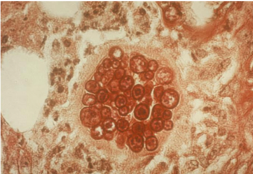
  • This shape shifter differs from others in that it does not shape shift by temperature but resembles other shape shifters in that it exists as a mold in the environment and a spherule (Image) inside the body.
  • Spherules are round, and contain endospores which are uninucleate and have walls and cytoplasmic inclusions
  • Can be mistaken for yeast if only endospores are seen (but endospores don’t bud like yeasts!)

Geography, Reservoir and Mode of Transmission
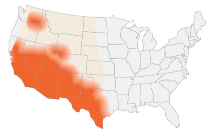
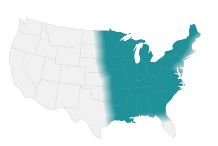
  • Endemic in southwest of the US and certain arid regions in South America
  • In the US, highest incidence in Arizona and California
  • Reservoir includes: soil (increased during dry periods after rain/storms/earthquakes or excavation work)
  • Mode of transmission: aerogenic
  • Race predilection: Filipino and African Americans at higher risk for disseminated disease (Board favorite!)

Clinical presentation

  • Two-thirds of individuals remain asymptomatic or develop self-limiting respiratory symptoms. When symptomatic → pulmonary involvement in >95% of cases(2)
  • The disease can spread hematogenously to the following extrapulmonary sites: skin, soft tissue, skeleton, CNS (both meninges and spinal cord), eyes, heart, liver, kidneys, and prostate(1)
  • Primary presentation often associated with skin findings (erythema nodosum, erythema multiforme) and rheumatological findings (myalgia, arthralgia)(2)
  • Eosinophilia is often present!
  • Unlike pulmonary nodules in histoplasmosis that often calcify, nodules in coccidiomycosis become small ‘grapeskin’ cavities, and cavities are often thin walled and can be associated with pleural effusions(3)

Diagnosis

Your friendly Infectious disease doctors will always ask for tissue, and if classic spherules are seen in histopathology or culture confirms growth, that’s a slam dunk diagnosis! But we understand that is not always feasible, so in addition to clinical history + presentation, in order of importance:

  1. Tissue/culture
  2. Serology (keeping in mind that it can be false negative very early on in the disease)

Culture:

*Please alert the microbiology lab if you suspect coccidiomycosis and are sending them cultures! (culture needs to be specially handled in the lab due to the risk occupational transmission/infection — just like all dimorphic fungi covered in this review series).

  • Confirms diagnosis, growth usually detected 4-5 days (4)

Histopathology:

  • Presence of endospore-containing spherules is diagnostic (see Image, Morphology)
  • Tissue eosinophilia may be present, and as endospores mature into spherules,
  • Granulomatous reaction predominates

Antigen detection:

  • Urinary antigen not widely used, however urinary histoplasma antigen test could be positive in 50% of cases(4)

Serology:

  • IgM usually becomes detectable within 1-3 weeks therefore negative serology early in the disease doesn’t rule out the disease
  • IgG becomes detectable anywhere between 3rd/4th week to several months after becoming infected. This usually reflects degree of infection and can be used to monitor the disease(4)
  • Increasing titers (or titers >1:32) may suggest dissemination(4)

Molecular methods:

  • No commercially available tests

Management(5)

Pulmonary disease:

  • Mild pulmonary disease: no treatment
  • Asymptomatic nodule/cavity: no treatment
  • Symptomatic chronic cavity: fluconazole/itraconazole (at least) for 12 months

Extra-pulmonary disease:

  • Soft tissue/bone: fluconazole/itraconazole (at least) for 12 months
    (*except for severe disease → Lipid Amphotericin B as an initial therapy for about 3 months)

References:

  1. Walsh TJ, Hayden RT, and Larone DH. Larone’s medically important fungi, 6th edition. ASM Press, 2018.
  2. Salzer HJF, Burchard G, Cornely OA, et al. Diagnosis and Management of Systemic Endemic Mycoses Causing Pulmonary Disease. Respiration. 2018; 96(3):283-301.
  3. Knox KS. Letter to the Editor: Perspective on Coccidioidomycosis and Histoplasmosis. Am J Resp Crit Care Medicine. 2014; 189(6):752-753.
  4. Saubolle MA, McKellar PP, and Sussland D. Epidemiologic, clinical, and diagnostic aspects of coccidiomycosis. J Clin Microbiol. 2007; 45(1):26-30.
  5. Galgiani JN, Ampel NM, Blair JE, et al. 2016 Infectious Diseases Society of America (IDSA) Clinical Practice Guideline for the Treatment of Coccidioidomycosis. Clin Infect Dis. 2016; 63(6):e112–e146
Blastomyces species

Learning about fungi is hard enough even for infectious disease fellows (Narrator: especially for infectious disease fellows). By the time you learn how to differentiate the yeasts from the molds, the fungi kingdom decides to throw you a curve ball: Enter the shape shifters into the game of fungi learning – the dimorphic fungi.

The Dimorphic fungi shape shift depending on the weather (literally). They exist as molds in the great outdoors (environmental temperatures) and yeasts in the great indoors (inside our bodies at body temperatures). Clinically, this also means you will see the yeast forms in a histopathology review of a tissue sample, and our friends in the microbiology lab can re-create the environmental factors to grow them out as mold forms in culture. So essentially, they also shape shift between the microbiology lab and the pathology department. (They are sneaky Fung(uy)i…).

If you haven’t read the first post on Histoplasma capsulatum, go read it now!

This is the 2nd post out of 6 and will focus on our second shapeshifter, Blastomyces dermatiditis.

CLICK HERE for a 2-page PDF handout of this information.

Morphology(1):

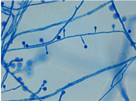
At 25°C-30°C (mold form): septate hyphae with short or long conidiophores where a pear-shaped conidia form at the apex of the conidiophore (has a lollipop-like appearance).
(Image A)

At 37°C (yeast form):
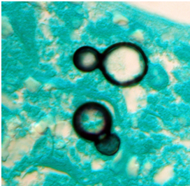
appear as yeast-like cells, thick walled and budding with a broad base
(Image B)

Geography, Reservoir and Mode of Transmission:

  • Endemic in North America (Ohio & Mississippi river valleys and the Great Lakes region)
  • Sporadic cases in Africa and India(2)
  • Reservoir includes: moist soil with decaying vegetative matter, decomposed wood
  • Mode of transmission: aerogenic, skin inoculation

Clinical presentation:

  • Incubation period: 30-45 days (3)
  • Spectrum of clinical disease:
    • Asymptomatic disease – occurs in ~50% of individuals
  • Acute pulmonary blastomycosis – resembles community-acquired pneumonia with variable presentation (infiltrates, consolidation +/- cavitation, reticulonodular patterns, small pleural effusions)(4)
    • Chronic pulmonary blastomycosis – can mimic presentation of TB, lung cancer, and histoplasmosis. Radiographic pattern often is described as alveolar or fibronodular infiltrations, mainly with an upper lobe distribution. Absence of mediastinal lymph node involvement in blastomycosis can distinguish it from Histoplasmosis.(4)
  • Extrapulmonary disease have been described in two-thirds of patients with chronic blastomycosis(3). Most frequent sites: skin, bones and genitourinary system.
  • Patients frequently present with cutaneous lesions without clinically active pulmonary disease.
  • CNS involvement is rare, except in immunocompromised hosts. As many as 40% of AIDs patients who have blastomycosis have CNS disease (mass lesions or meningitis)(5).

Diagnosis:

Your friendly Infectious disease doctors will alwaysask for tissue, and if classic broad-based budding yeast are appreciated in histopathology, that’s a slam dunk diagnosis! But we understand that is not always feasible, so in addition to clinical history + presentation, in order of importance:

  1. Tissue (histopathology, faster visualization, culture will have a long incubation time)
  2. Urine Antigen detection (keeping in mind issues with cross-reactivity as outlined below)

Culture:

*Please alert the microbiology lab if you suspect Blastomycosis and are sending them cultures! (culture needs to be specially handled in the lab due to risk occupational transmission/infection (just like all dimorphic fungi covered in this review series).

  • Most sensitive method, however long incubation time
  • For cutaneous lesions, important to obtain specimen from active leading edge

Histopathology:

  • Forms non-caseating granulomas
  • Classic appearance in clinical samples: Broad-based budding yeast (Image B, morphology)

Antigen detection:

High sensitivity for urine detection (93%) in largest published evaluation(2) but low specificity (79%) due to cross reactivity with Histoplasmosis, Paracoccidioidomycosis and Talaromycosis (previously known as penicilliosis)

Serology:

  • No role in diagnosis because of poor sensitivity and high cross-reactivity(4)

Molecular methods:

  • No commercially available tests

Management(3):

Pulmonary disease

  • Mild to moderate: Itraconazole 6-12 months
  • Moderate to severe: Lipid Amphotericin B for 1-2 weeks followed by →  Itraconazole for 6-12 months

Disseminated disease

  • Mild to moderate: Itraconazole for 6-12 months
  • Moderate to severe: Lipid Amphotericin B for 1-2 weeks followed by →  Itraconazole for 12 months

References:
1. Walsh TJ, Hayden RT, Larone DH. Larone’s medically important fungi, 6th edition, ASM press, 2018.
2. Saccente M and Woods GL. Clinical and laboratory update on blastomycosis. Clin Microbiol Rev. 2010;23:367–381.
3. Chapman SW, Dismukes WE, Proia LA, Bradsher RW, Pappas PG, Threlkeld MG, et al. Clinical practice guidelines for the management of blastomycosis: 2008 update by the Infectious Diseases Society of America. Clin Infect Dis. 2008 Jun 15;46(12):1801-12
4. Salzer HJF, Burchard G, Cornely OA, et al. Diagnosis and management of systemic endemic mycoses causing pulmonary disease. Respiration; 2018;96:283–301.
5. Pappas PG, Pottage JC, Powderly WG, et al. Blastomycosis in patients with the acquired immunodeficiency syndrome. Ann Intern Med;1992:116

Histoplasma Capsulatum

Learning about fungi is hard enough even for infectious disease fellows (Narrator: especially for infectious disease fellows). By the time you learn how to differentiate the yeasts from the molds, the fungi kingdom decides to throw you a curve ball: Enter the shape shifters into the game of fungi learning – the dimorphic fungi.

The Dimorphic fungi shape shift depending on the weather (literally). They exist as molds in the great outdoors (environmental temperatures) and yeasts in the great indoors (inside our bodies at body temperatures). Clinically, this also means you will see the yeast forms in a histopathology review of a tissue sample, and our friends in the microbiology lab can re-create the environmental factors to grow them out as mold forms in culture. So essentially, they also shape shift between the microbiology lab and the pathology department. (They are sneaky Fung(uy)i…)

This is the first post out of 6 and will focus on our first shapeshifter, Histoplasma capsulatum.

CLICK HERE for a 2-page PDF handout of this information.

Morphology:(3)

  • At 25°C-30°C (mold form):Young cultures – septate hyphae with smooth or spiny microconidia
  • Older cultures (several weeks old) – large, thick walled round macroconidia with knobby projections (Image)
  • At 37°C (yeast form): small, round budding cells 

Geography, Reservoir and Mode of Transmission:

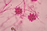
  • Histoplasma has a world-wide distribution(4), but is mostly endemic in the Americas (Central/Eastern United States & Central and South America)
  • Reservoir includes: soil, areas of construction, animal droppings (i.e. bats – a board favorite!), and caves (another board favorite)
  • Mode of transmission: aerogenic

Clinical presentation:

  • Known as the ‘syphilis’ of the fungal world because it’s a great imitator, particularly of TB(4).
  • Disease presentation/severity depends on size of inoculum & immune status
    • Immunocompetent hosts: usually asymptomatic/self- limiting
    • Immunocompromised hosts: often progressive/severe/disseminated
  • Can present as:
    • Acute pulmonary disease: Either as diffuse or localized infiltrates +/- mediastinal lymphadenopathy
    • Chronic pulmonary disease: Cavitary lesions/nodules
    • Mediastinal disease: Mediastinal granulomas and fibrosis

Diagnosis:

1. Culture:

  • *Please alert the microbiology lab if you suspect histoplasmosis and are sending them cultures! (culture needs to be specially handled in the lab due to risk occupational transmission/infection (just like all dimorphic fungi covered in this review).
  • Sensitivity of both tissue and blood cultures depend on the presentation (pulmonary vs. disseminated), immune status and burden of disease(5)
  • Disseminated disease → ~74% will have positive cultures(6)
  • Pulmonary disease → ~42% will have positive cultures(6)
  • HIV/AIDS patients:
    → ~ 90% of respiratory cultures will be positive(7)
    →~50% of blood cultures will be positive(7)

2. Histopathology:

  • Appear as yeast form, predominantly phagocytosed within macrophages and histiocytes
  • Presence in tissue supports diagnosis, although does not necessarily indicate active infection (could be detected in non-active granulomas for years)
  • Characteristic pathology feature is the presence of granulomas (caseating or non-caseating)(6)

3. Antigen detection:

  • Preferred method of testing: rapid testing + non-invasive + highly sensitive.
  • Sensitivity: urinary antigen > serum antigen (across all spectrum of clinical presentations of histoplasmosis)(9)
  • Histoplasma serum antigen (MiraVista© EIA) have highest sensitivity in disseminated disease (91.8%) >chronic pulmonary disease (87.5%) >acute pulmonary disease (83%) >subacute histoplasmosis (30%)(8)
  • In HIV/AIDS patients with disseminated disease, Histoplasma antigen can be detected in 95% of cases
  • Mediastinal involvement in histoplasmosis (mediastinal granuloma, mediastinitis) doesn’t usually result in positive antigen testing
  • Histoplasma antigen can cross react with all the dimorphic fungi covered in this review series (less commonly for coccidioides spp.)

4. Serology:

  • Antibodies take 4-8 weeks to become detectable therefore not useful for acute diagnosis but can be helpful for subacute and chronic forms of the disease
  • Titers usually decrease with disease resolution, but slowly so titers cannot be used to monitor for treatment response
  • Immunocompromised patients, particularly those with humoral defects, might not mount an antibody response so serology testing isn’t as useful.

5. Molecular methods:

  • No currently FDA approved molecular assay for H. capsulatum for clinical use.
  • PCR assays available in reference labs but are not yet standardized

Management(12):

Clinical presentationMild/ModerateModerate/SevereChronic
Pulmonary<4weeks: none
>4weeks: itraconazole for 6-12 months
Lipid amphotericin B for 1-2 weeks followed by itraconazole for 12 weeksItraconazole for 12 months
DisseminatedItraconazole for 12 monthsLipid amphotericin B for 1-2 weeks followed by itraconazole for 12 monthsN/A

References:
1. Climate change: the role of the infectious disease community. Lancet Infect Dis. 2017; 17:1219.
2. Greer A, Ng V, and Fisman D. Climate change and infectious diseases in North America: the road ahead. CMAJ. 2008; 178:715–722.
3. Walsh, TJ, Hayden, RT, and Larone, DH. Larone’s medically important fungi, 6th edition, ASM press, 2018.
4. Queiroz-Telles F, Fahal AH, Falci DR, et al. Neglected endemic mycoses. Lancet Infect Dis. 2017;17:e367–e377.
5. Azar MM and Hage CA. Laboratory Diagnostics for Histoplasmosis. J Clin Microbiol. 2017; 55:1612–1620.
6. Hage CA, Azar MM, Bahr N, Loyd J, and Wheat LJ. Histoplasmosis: up-to-date evidence-based approach to diagnosis and management. Semin Respir Crit Care Med. 2015; 36:729–745. 
7. Kauffman CA. Histoplasmosis: a clinical and laboratory update. Clin Microbiol Rev. 2007;20:115–132.
8. Hage CA, Ribes JA, Wengenack NL, et al. A multicenter evaluation of tests for diagnosis of histoplasmosis. Clin Infect Dis. 2011;53:448–454. 
9. Wheat LJ and Kauffman CA. Histoplasmosis. Infect Dis Clin North Am. 2003;17:1–19.
10. Swartzentruber S, Rhodes L, Kurkjian K, et al. Diagnosis of acute pulmonary histoplasmosis by antigen detection. Clin Infect Dis. 2009; 49:1878–1882. 
11. Saccente M and Woods GL. Clinical and laboratory update on blastomycosis. Clin Microbiol Rev. 2010;23:367–381.
12. Wheat LJ, Freifeld AG, Kleiman MB, et al; Infectious Diseases Society of America. Clinical practice guidelines for the management of patients with histoplasmosis: 2007 update by the Infectious Diseases Society of America. Clin Infect Dis. 2007;45:807–825.

The Shape Shifters: Dimorphic fungi

Learning about fungi is hard enough even for infectious disease fellows (Narrator: especially for infectious disease fellows). By the time you learn how to differentiate the yeasts from the molds, the fungi kingdom decides to throw you a curve ball: Enter the shape shifters into the game of fungi learning – the dimorphic fungi.

The Dimorphic fungi shape shift depending on the weather (literally). They exist as molds in the great outdoors (environmental temperatures) and yeasts in the great indoors (inside our bodies at body temperatures). Clinically, this also means you will see the yeast forms in a histopathology review of a tissue sample, and our friends in the microbiology lab can re-create the environmental factors to grow them out as mold forms in culture. So essentially, they also shape shift between the microbiology lab and the pathology department. (They are sneaky Fung(uy)i…)

Some of the clinically relevant dimorphic fungi have a predilection for geographical location (endemic mycoses), and therefore are very popular in board exams to the dismay (or joy, after this review series?) of medical trainees.

#ClimateChangeIsReal isn’t just pertinent in the political arena, but also for these endemic fungi. The grave consequences of climate change might change and expand the geographical distribution(1,2) of these fungi and therefore result in more catch-up learning on our end. This is almost akin to learning the constant re-classification and re-naming of the fungi kingdom (thanks, no thanks taxonomists…)

In this review series, I will go over the endemic fungi in a ‘high yield’ approach that will hopefully be pertinent for both shelf exams/boards and clinical practice.

I’ve also purposefully made it a two-pager/per fungi review (or 1 pager if you print it double-sided, #SaveTheTrees). We will be providing PDF links with every Fungi review. This will be an easy reference for a pocketbook, handouts to print to teach your medical students or if you want to flex your knowledge of endemic fungi during rounds (All win-win-win situations!)

The profile of each shape shifter will be released every Friday in the spirit of #FungalFriday. The dimorphic fungi that will be covered during the #ShapeShifterSeries include:

  • Histoplasmosis
  • Blastomycosis
  • Coccidioidomycosis
  • Talaromycosis
  • Paracoccidiomycosis
  • Sporotrichosis

Our First Shape shifter in the series to be released this coming #FungalFriday will be Histoplasmosis, aka the Ohio valley disease/Cave disease. What does Ohio or caves for the matter have to do with this Fungus? Find out more this coming Friday!

Fatima Al Dhaheri, MBBS
The Fung(uy)i squad

References:
1. The Lancet Infectious Diseases. Climate change: the role of the infectious disease community. Lancet Infect Dis. 2017;17:1219.
2. Greer A, Ng V, Fisman D. Climate change and infectious diseases in North America: the road ahead. CMAJ. 2008;178:715–722.


Clostridium difficile

In light of the recently published IDSA guidelines on C. difficile, I thought I would write up a summary of the guidelines as well as provide some of the background microbiology of the organism for review.

Structure

  • obligate anaerobes
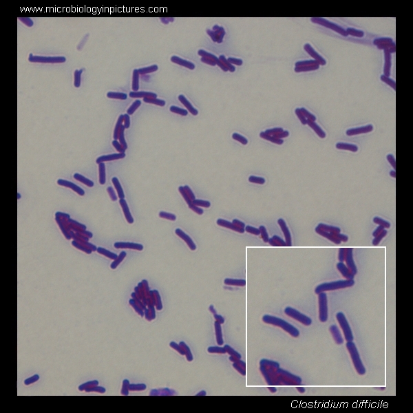
  • gram-positive bacilli
    – form spores (= dormant, non-reproductive structure that the bacteria can reduce itself to in order to survive for extended periods of time in extreme conditions)
  • produce toxins (toxin A and toxin B) that cause disease


clostridium-difficile gram stain

Environment

  • animal and human feces
  • soil
  • sewage


Mechanism of pathogenicity

*Not all strains of C. difficile are pathogenic – only the ones who produce toxins can cause C. difficile disease

Pathogenesis:

Transmission occurs with ingestion of spores via the fecal-oral route ⇒ spores activate in the colon to replicating bacteria ⇒ bacteria release toxins ⇒ toxins cause breakdown of the colon cells’ cytoskeleton framework ⇒ apoptosis ⇒ breakdown of the mucosal wall ⇒ DIARRHEA!


Risk factors for acquiring C. difficile:

  1. ANTIBIOTIC EXPOSURE
  2. Exposure to healthcare facilities
  3. Age and immunosuppression
  4. ?Gastric acid suppression (use of proton pump inhibitors or H2 receptor blockers)
    — the evidence-based-medicine jury is still out on this one


Clinical features

  • symptoms can develop during antibiotic treatment or up to 6 weeks after the course of antibiotics has been finished
  • patients can also become infected even without exposure to antibiotics (both in the healthcare setting but also in the community setting)
  • carrier state = a patient who is colonized with C. difficile but is currently asymptomatic

1.Symptoms and physical exam signs:

  • Non-bloody, WATERY DIARRHEA (≥ 3 loose stools in 24 hours)
    *occasionally patients can develop ileus with severe infection which will not result in diarrhea but rather lack of bowel movements
  • Abdominal pain/cramping
  • Fever and chills
  • Abdominal distention/tenderness
  • Nausea/anorexia

2.Laboratory findings:

  • High white blood count (occasionally precedes the diarrhea by 1-2 days)
  • Elevated creatinine
  • Elevated lactate and low albumin (in fulminant cases)


Pseudomembranous colitis = inflammation of the colon causing elevated white and yellow-colored plaques to form and coalesce together to create a pseudomembrane on the colon wall that can be seen by colonoscopy.


Diagnostics

  1. When to test: when patient has new onset, ≥ 3 unformed stools that cannot be explained by another cause (i.e. laxative use)
  2. Options for testing:

* C. difficile can be grown in culture, but anaerobes take a while to grow and it would not provide an answer as to whether the strain is toxigenic (i.e. produces toxin) or not, so it is not commonly used for clinical diagnostic purposes.

What it is Advantages Disadvantages
Toxin EIA assay Antibody assay that detect toxins High specificity (>84%) Low sensitivity (31-99%)
GDH assay Detects GDH (an enzyme produced by C. difficile) High NPV (>99%)

Quick turn-around

Cannot distinguish toxigenic vs. non-toxigenic C. difficile strains
NAAT/PCR PCR method that detects toxin production gene High NPV (>99%)

Quick turn-around

Poor specificity and PPV

*Changes depending on whom specimens are collected on (low suspicion vs. high suspicion)

EIA, enzyme immunoassay; GDH, glutamate dehydrogenase; NPV, negative predictive value; PPV, positive predictive value; NAAT, nucleic acid amplification test; PCR, polymerase chain reaction.

Many healthcare facilities are currently doing only PCR testing. It’s highly sensitive and the results return quickly (usually within 24 hours).

The problem: this practice is yielding a lot of false positives (patients who are carriers but do not truly have an active infection) which ⇒ over-treatment ⇒ patient discomfort, potential side effects, infection control consequences for the hospital, and extra costs.

Why: This is thought to be due to the fact that a lot of tests are sent inappropriately (on patients that have diarrhea but no other evidence of infection such as leukocytosis, AKI, abdominal pain, fever, etc.)

Solution (as proposed by IDSA guidelines):

  1. A multiple step algorithm:
  • GDH assay + EIA assay
  • GDH assay + EIA assay with NAAT as a tiebreaker
  • NAAT + EIA assay

                  OR

  1. We can agree to be more mindful of when we send the test (when the pre-test probability is high) and continue to use the NAAT/PCR method alone.

Bottom line: Many hospitals are switching over to the two-step testing method for multiple reasons:

  • behavior change is difficult to implement and sustain
  • provides more accurate incidence of nosocomial-acquired infections in the hospital

WHEN YOU THINK OF SENDING A C. DIFFICILE TEST, ask yourself:

  1. Does this patient have an unexplained fever, leukocytosis, or new abdominal pain/distention, in addition to the diarrhea (or in presence of ileus)?
    if yes ⇒ send the test
  1. If not, is there another explanation for the diarrhea?
    (i.e. laxatives, new medications (especially antibiotics), part of already known illness, etc.)
    if yes ⇒ consider removing the potential cause (if possible) and re-evaluate or monitor for worsening symptoms
    if not ⇒ send the test


Treatment

The IDSA has a really great table to reference when choosing treatment options for your C. difficile infected patient.

***PO Metronidazole is no longer the 1st-line agent for C. diff infection treatment***

C.diff treatment chart

                                                                                                            McDonald et al. CID 2018


Recurrence

Recurrence of C. difficile infection = reappearance of symptoms within 2-8 weeks after completion of therapy

  • up to 25% of patients will experience a recurrence
  • once patient had one recurrence, they are at higher risk for future recurrences


TAKE-HOME POINTS:

  • The MAJOR risk factor for C. difficile infection is ANTIBIOTIC EXPOSURE
    ⇒ DO NOT give antibiotics to those who do not truly need them
  • Symptoms/signs include watery diarrhea, abdominal cramping/pain, and elevated WBC and creatinine
  • C. difficile infection CAN cause ileus (i.e. no diarrhea)
  • Only send test when you have a high pre-test probability to avoid false positives
  • Metronidazole is NO LONGER recommended for treatment of C. difficile


Got questions? Disagree? Leave your comments below!


References

  1. McDonald LC, Gerding DN, Johnson S, et al. Clinical Practice Guidelines for Clostridium difficile Infection in Adults and Children: 2017 Update by the Infectious Diseases Society of America (IDSA) and Society for Healthcare Epidemiology of America (SHEA). Clin Infect Dis. 2018. 66(7):1-48. [PMID: 29562266]
  2. Jorgensen JH, Pfaller MA, Carroll KC, et al. Manual of Clinical Microbiology, Eleventh Edition.
  3. Lamont JT. (2018). Clostridium difficile infection in adults: Epidemiology, microbiology, and pathophysiology. In Baron EL, ed. UpToDate. Waltham, Mass.: UpToDate, 2018. [https://www.uptodate.com/contents/clostridium-difficile-infection-in-adults-epidemiology-microbiology-and-pathophysiology]. Accessed May 25, 2018.
  4. Lamont JT, Kelly CP, and Bakken JS. Clostridium difficile infection in adults: Clinical manifestations and diagnosis. In Baron EL, ed. UpToDate. Waltham, Mass.: UpToDate, 2018. [https://www.uptodate.com/contents/clostridium-difficile-infection-in-adults-clinical-manifestations-and-diagnosis]. Accessed May 25, 2018.
  5. Kelly CP, Lamont JT, and Bakken JS. Clostridium difficile infection in adults: Treatment and prevention. In Baron EL, ed. UpToDate. Waltham, Mass.: UpToDate, 2018. [https://www.uptodate.com/contents/clostridium-difficile-infection-in-adults-treatment-and-prevention]. Accessed May 25, 2018.

Blood culture contaminants

There are some bacteria out there that usually don’t cause disease. They tend to just hang out and not cause any harm. When we see them in the bloodstream, they often are contaminants, meaning they were introduced during collection of the sample, and are not truly in the bloodstream. Figuring out which ones are contaminants and which ones are not can be tricky. Some are almost always (see * below) contaminants while others are NEVER contaminants. Some can be a contaminant or an infection, like streptococcus viridans group.

*Important to note: any organism can cause an infection in the right context, even those who are usually deemed as contaminants. If you are unsure whether a true infection is present, it’s always best to call an infectious disease specialist to assist with management.

Contaminant =  growth of bacteria in the blood culture bottle that were not present in the patient’s bloodstream and thus introduced during the collection of the sample (Dawson et al.)

NEVER assumed to be contaminants:

Staphylococcus aureus
Streptococcus pneumoniae
Group A/B streptococcus
Listeria monocytogenes
Neisseria meningitidis
Gram negative bacilli
Fungus (yeast or mold)
…Amongst many other numerous organisms that will take too long to mention in a list

Common blood contaminants:

Coagulase-negative staphylococci
Propionibacterium acnes
Micrococcus spp.
Corynebacterium spp.
Bacillus spp. (*except Bacillus anthracis which causes anthrax!!)

What if multiple bacteria are growing?!

  • multiple growth of bacteria can suggest contamination.
  • however, it again depends on the pathogens involved. If all are skin flora, then probably contaminant. If any of the aforementioned ‘USUALLY NOT CONTAMINANTS’ are present, then it is not a contaminant!


Where do contaminants come from:

  1. Patient’s skin
  2. Equipment used to obtain sample and transfer it to culture bottle
  3. Provider’s skin
  4. General environment


Factors to consider when deciding whether culture
is contaminant or not:

  1. Patient’s clinical status
    • are there any signs or symptoms to suggest infection with this microbe?
    • Example:
      • You would not expect a patient with pneumonia to grow CoNS in their blood.
      • However, you would take CoNS seriously in a patient with recent valve replacement and fevers.
  1. Microbiology of the species – see above
  2. Time to positivity of blood culture – studies have shown that the average time of positivity of true infections is ~12 hours vs. ~24 hours for contaminants
  3. Inoculum of the isolate – how much growth is there?
    • If only 1 out of 4 blood culture bottles are positive –> MORE LIKELY contaminant
    • If >1 out of 4 blood culture bottles are positive –> true pathogen
  1. Any foreign material? – Contaminants become pathogens when they infect hardware and prosthetic valves/grafts. If those are present and there is a possibility they are infected, would not disregard any pathogen as a contaminant.
  2. Patient’s response to antibiotics and isolate’s susceptibility pattern – clues to whether an isolate is a contaminant or a true infection
    • Contaminant: patient does not respond to antibiotics that treat the blood culture isolate
    • True pathogen: patient DOES respond to antibiotics that treat the blood culture isolate
    • Contaminant: patient responds to antibiotics despite blood culture isolate resistant
    • True pathogen: patient does not respond to antibiotics and isolate is resistant


FUN FACT: A retrospective review looked at 626 blood cultures and discovered that by 48 hours, 98% of aerobic gram-positive and gram-negative bloodstream infections were identified.

*This shows that unless there is a high suspicion for anaerobe growth, antibiotics can be de-escalated at 48 hours if there is no growth. (Pardo, J., Klinker, K.P, et al. 2014. Time to positivity of blood cultures supports antibiotic de-escalation at 48 hours. Annals of Pharmacology. 48 (1): 33-40).)

TAKE HOME POINTS:

  • Microbes known to be common contaminants CAN cause disease in certain circumstances
  • Always repeat blood culture when first one is positive for microbes
  • If you’re not sure if it’s a contaminant or not ⇒ call ID
    (it’s always better to double check rather than to miss a true infection)


References:

1. Dawson, S. 2014. Blood culture contaminants. Journal of Hospital Infection; 87, 1-10. DOI: 10.1016/j.jhin.2014.02.009
2. Pardo, J., Klinker, K.P, et al. 2014. Time to positivity of blood cultures supports antibiotic de-escalation at 48 hours. Annals of Pharmacology. 48 (1): 33-40).
DOI: 10.1177/1060028013511229
3. Hossain, B., Islam, M.S., et al. 2016. Understanding bacterial isolates in blood culture and approaches used to define bacteria as contaminants: a literature review. Pediatric Infectious Disease Journal. 35(5): S45-51.
DOI: 10.1097/INF.0000000000001106
4. Pien, B.C., Sundaram, P., and et al. 2010. The clinical and prognostic importance of positive blood cultures in adults. American Journal of Medicine. 123 (819-828).
DOI: 10.1016/j.amjmed.2010.03.021

SPICE organisms

First topic at hand is SPICE organisms. These are the organisms that appear to be sensitive to many antibiotics, but once they are exposed to certain antibiotics (ex. 3rd generation cephalosporins), they quickly develop resistance to them.

SPICE stands for:

S: Serratia spp.

P: Providencia

I: “indole-positive” Proteus spp. (this includes: P. vulgaris) *NOT P.mirabilis

C: Citrobacter spp.

E: Enterobacter spp.

*There are other, less known bacteria included in this group (Cronobacter, Edwardsiella, Hafnia, Morganella, Aeromonas)

*[Organisms like Pseudomonas and Acinetobacter produce AmpC gene normally – which is why they have intrinsic resistance to 3rd generation cephalosporins and do not technically fall into the AmpC inducer SPICE group.]

The SPICE pathogens can be induced to produce an AmpC beta-lactamase gene that encodes an enzyme that cleaves the beta-lactam group in the antibiotic and renders it inactive.

This gene may not be detected initially (low level of expression of the gene) but may appear (induced to express higher levels of gene) after a period of exposure to beta-lactam antibiotics.

(Clinical translation: Initially they will appear susceptible to beta-lactams, but eventually will develop resistance to them)

Once beta-lactam is removed, the AmpC gene production is reduced once more and the pathogens will appear sensitive to 3rd generation cephalosporins and penicillins again. .

Resistance develops anywhere from 24h to 2-3 weeks.

Clinical relevance:

  • If the course of antibiotics is short or if the antibiotic can easily overcome the MIC concentration needed for bacterial killing, then the risk of inducing AmpC gene production is low
    • Clinical examples (~<1 week duration of antibiotics):
      • UTI
      • Pneumonia
  • Short course for intra-abdominal infectionHowever, this becomes an issue in areas where antibiotics have difficulty penetrating (because it is less likely to overcome the MIC concentration needed) or when antibiotics are needed to be given over a longer period of time.
    • Clinical examples:
      • Endocarditis
      • Bacteremia
      • Osteomyelitis
      • Septic arthritis
      • Abscesses

Antibiotics to avoid:

  • Penicillin class (including piperacillin-tazobactam)
  • Most cephalosporins (1st, 2nd, and 3rd generation)

Antibiotics to use:

  • 4th generation cephalosporins (i.e. cefepime at higher doses, q8h)
  • Carbapenems
  • Aminoglycosides
  • Fluoroquinolones

TAKE-HOME POINTS:

  1. Remember the members of the SPICE group
  2. You may be successful in treating an infection in short courses of therapy or in infections where antibiotic penetration is high. But in patients with bacteremia, bone, joint, or valve infections – strongly consider 4th generation cephalosporin or a carbapenem.

Have a question, comment, or a suggestion for a future blog post? Post your comment below!

References:

  • http://m.antimicrobialstewardship.com/clinical_summaries/index.php?page=esbl_and_spice
  • Jacoby, G.A. AmpC beta-lactamases. Clinical Microbiology Review. 2009. 22(1):161-182. doi: 10.1128/CMR.00036-08
  • Harris, P.N.A, and Ferguson, J.K. Antibiotic therapy for inducible AmpC beta-lactamase-producing Gram-negative bacilli: what are the lternatives to carbapenems, quinolones, and aminoglycosides? 2012. International Journal of Antimicrobial Agents, 40: 297-305.