Tag Archives: #infectious diseases

COVID-19 Ultimate Resource List

There’s so much information about the novel coronavirus COVID-19 on the internet that it is hard to keep up with the onslaught of information. We wanted to compile the ultimate list of reputable resources for clinical providers to access when they need to, at a moment’s time.

Since information on COVID-19 is rapidly changing, these resources are not specific papers or blog posts, but rather websites that are maintaining up to date information on epidemiology, pathophysiology, and management. Resources span all types, including website behemoths like the WHO and CDC, as well as twitter accounts for people who get their news from social media.

Since this is an ultimate list but we don’t pretend to have ultimate knowledge of all resources, please send us resources that we may have missed and we will add them on here! We also acknowledge that this list is U.S. centric because we are from the States, but we would love input on resources for other countries so we can make this a more international list.

For the most up to date information:

Epidemiology:

Academic:

Clinical/Practical Management:

  • University of Washington – the holy grail of practical documents developed by people who are currently battling a COVID-19 outbreak. All documents can be utilized by others and adapted for their own institutional protocols.
  • University of Kentucky – have assessment, triage, and testing algorithms for use
  • University of Nebraska Medical Center – fantastic protocols on donning on/off PPE, videos, as well as other in-hospital quarantine protocols
  • IDStewardship – a very thorough, but easily read overview of potential treatment options for COVID-19 and how/when to use them
  • University of Liverpool – charts on drug drug interactions with experimental therapies for COVID-19
  • CIDRAP – focuses on the health policy aspect of COVID-19 news
  • Society of Critical Care Medicine – online training modules for disaster medicine, resource availability in U.S., and other fantastic resources for front-line clinical providers

Immunocompromised hosts:

Clinical Trials:

  • ClinicalTrials.gov – a currently-recruiting randomized controlled trial of remdesivir for COVID-19 treatment
  • CURE ID –  an FDA and NIH supported website where you can report and see new uses of existing drugs for difficult-to-treat infectious diseases. Download their mobile app too!

Blogs:

Podcasts:

  • TWiV – a podcast by virologists discussing virology; with recent episodes on COVID-19
  • IDSA Podcast – has weekly podcast series “Coronavirus: What’s happening now?”
  • JAMA Clinical Reviews – recent episodes on COVID-19 from various different viewpoints
  • NEJM Interviews – hosts short and to the point interviews with world experts
  • EMCrit Podcast – EMCrit also has a podcast! Run by Scott Weingart, for those who don’t have time to read.
  • FOAMCast (for the interim also COVIDCast) – an EM podcast run by Lauren Westafer and Jeremy Faust, they provide DAILY high yield updates on COVID-19 literature
  • This Podcast Will Kill You – Two epidemiologists (both named Erin) provide you with an entertaining summary of new COVID-19 knowledge, alongside a recipe for a Quarantini and a Placeborita.

Twitter accounts:

Graphics:

Graphics Reuters – for the graphic enthusiasts out there
New York Times Coronavirus Maps – for the graphic enthusiasts out there

That’s it for now! Let us know if we missed any great resources!

Thank you for all the work that all of you do. Stay safe!

This list was compiled by Milana Bogorodskaya, Fatima Al-Dhaheri, and Ahmed Abdul Azim.

Coccidioides species

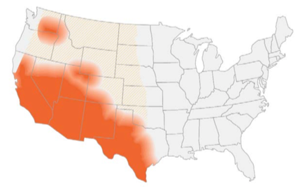
Disease: Coccidioidomycosis

Alias: Valley fever, San Joaquin fever

Learning about fungi is hard enough even for infectious disease fellows (Narrator: especially for infectious disease fellows). By the time you learn how to differentiate the yeasts from the molds, the fungi kingdom decides to throw you a curve ball: Enter the shape shifters into the game of fungi learning – the dimorphic fungi.

The Dimorphic fungi shape shift depending on the weather (literally). They exist as molds in the great outdoors (environmental temperatures) and yeasts in the great indoors (inside our bodies at body temperatures). Clinically, this also means you will see the yeast forms in a histopathology review of a tissue sample, and our friends in the microbiology lab can re-create the environmental factors to grow them out as mold forms in culture. So essentially, they also shape shift between the microbiology lab and the pathology department. (They are sneaky Fung(uy)i…).

If you haven’t read the posts on Histoplasma capsulatum or Blastomyces species, go read it now!

This is the 3rd post out of 6 and will focus on our third shapeshifter, Coccidioides species.

CLICK HERE for a 2-page PDF handout of this information.

Morphology(1):

  • This shape shifter differs from others in that it does not shape shift by temperature but resembles other shape shifters in that it exists as a mold in the environment and a spherule (Image) inside the body.
  • Spherules are round, and contain endospores which are uninucleate and have walls and cytoplasmic inclusions
  • Can be mistaken for yeast if only endospores are seen (but endospores don’t bud like yeasts!)

Geography, Reservoir and Mode of Transmission

  • Endemic in southwest of the US and certain arid regions in South America
  • In the US, highest incidence in Arizona and California
  • Reservoir includes: soil (increased during dry periods after rain/storms/earthquakes or excavation work)
  • Mode of transmission: aerogenic
  • Race predilection: Filipino and African Americans at higher risk for disseminated disease (Board favorite!)

Clinical presentation

  • Two-thirds of individuals remain asymptomatic or develop self-limiting respiratory symptoms. When symptomatic → pulmonary involvement in >95% of cases(2)
  • The disease can spread hematogenously to the following extrapulmonary sites: skin, soft tissue, skeleton, CNS (both meninges and spinal cord), eyes, heart, liver, kidneys, and prostate(1)
  • Primary presentation often associated with skin findings (erythema nodosum, erythema multiforme) and rheumatological findings (myalgia, arthralgia)(2)
  • Eosinophilia is often present!
  • Unlike pulmonary nodules in histoplasmosis that often calcify, nodules in coccidiomycosis become small ‘grapeskin’ cavities, and cavities are often thin walled and can be associated with pleural effusions(3)

Diagnosis

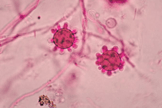
Your friendly Infectious disease doctors will always ask for tissue, and if classic spherules are seen in histopathology or culture confirms growth, that’s a slam dunk diagnosis! But we understand that is not always feasible, so in addition to clinical history + presentation, in order of importance:

  1. Tissue/culture
  2. Serology (keeping in mind that it can be false negative very early on in the disease)

Culture:

*Please alert the microbiology lab if you suspect coccidiomycosis and are sending them cultures! (culture needs to be specially handled in the lab due to the risk occupational transmission/infection — just like all dimorphic fungi covered in this review series).

  • Confirms diagnosis, growth usually detected 4-5 days (4)

Histopathology:

  • Presence of endospore-containing spherules is diagnostic (see Image, Morphology)
  • Tissue eosinophilia may be present, and as endospores mature into spherules,
  • Granulomatous reaction predominates

Antigen detection:

  • Urinary antigen not widely used, however urinary histoplasma antigen test could be positive in 50% of cases(4)

Serology:

  • IgM usually becomes detectable within 1-3 weeks therefore negative serology early in the disease doesn’t rule out the disease
  • IgG becomes detectable anywhere between 3rd/4th week to several months after becoming infected. This usually reflects degree of infection and can be used to monitor the disease(4)
  • Increasing titers (or titers >1:32) may suggest dissemination(4)

Molecular methods:

  • No commercially available tests

Management(5)

Pulmonary disease:

  • Mild pulmonary disease: no treatment
  • Asymptomatic nodule/cavity: no treatment
  • Symptomatic chronic cavity: fluconazole/itraconazole (at least) for 12 months

Extra-pulmonary disease:

  • Soft tissue/bone: fluconazole/itraconazole (at least) for 12 months
    (*except for severe disease → Lipid Amphotericin B as an initial therapy for about 3 months)

References:

  1. Walsh TJ, Hayden RT, and Larone DH. Larone’s medically important fungi, 6th edition. ASM Press, 2018.
  2. Salzer HJF, Burchard G, Cornely OA, et al. Diagnosis and Management of Systemic Endemic Mycoses Causing Pulmonary Disease. Respiration. 2018; 96(3):283-301.
  3. Knox KS. Letter to the Editor: Perspective on Coccidioidomycosis and Histoplasmosis. Am J Resp Crit Care Medicine. 2014; 189(6):752-753.
  4. Saubolle MA, McKellar PP, and Sussland D. Epidemiologic, clinical, and diagnostic aspects of coccidiomycosis. J Clin Microbiol. 2007; 45(1):26-30.
  5. Galgiani JN, Ampel NM, Blair JE, et al. 2016 Infectious Diseases Society of America (IDSA) Clinical Practice Guideline for the Treatment of Coccidioidomycosis. Clin Infect Dis. 2016; 63(6):e112–e146

Histoplasma Capsulatum

Learning about fungi is hard enough even for infectious disease fellows (Narrator: especially for infectious disease fellows). By the time you learn how to differentiate the yeasts from the molds, the fungi kingdom decides to throw you a curve ball: Enter the shape shifters into the game of fungi learning – the dimorphic fungi.

The Dimorphic fungi shape shift depending on the weather (literally). They exist as molds in the great outdoors (environmental temperatures) and yeasts in the great indoors (inside our bodies at body temperatures). Clinically, this also means you will see the yeast forms in a histopathology review of a tissue sample, and our friends in the microbiology lab can re-create the environmental factors to grow them out as mold forms in culture. So essentially, they also shape shift between the microbiology lab and the pathology department. (They are sneaky Fung(uy)i…)

This is the first post out of 6 and will focus on our first shapeshifter, Histoplasma capsulatum.

CLICK HERE for a 2-page PDF handout of this information.

Morphology:(3)

  • At 25°C-30°C (mold form):Young cultures – septate hyphae with smooth or spiny microconidia
  • Older cultures (several weeks old) – large, thick walled round macroconidia with knobby projections (Image)
  • At 37°C (yeast form): small, round budding cells 

Geography, Reservoir and Mode of Transmission:

  • Histoplasma has a world-wide distribution(4), but is mostly endemic in the Americas (Central/Eastern United States & Central and South America)
  • Reservoir includes: soil, areas of construction, animal droppings (i.e. bats – a board favorite!), and caves (another board favorite)
  • Mode of transmission: aerogenic

Clinical presentation:

  • Known as the ‘syphilis’ of the fungal world because it’s a great imitator, particularly of TB(4).
  • Disease presentation/severity depends on size of inoculum & immune status
    • Immunocompetent hosts: usually asymptomatic/self- limiting
    • Immunocompromised hosts: often progressive/severe/disseminated
  • Can present as:
    • Acute pulmonary disease: Either as diffuse or localized infiltrates +/- mediastinal lymphadenopathy
    • Chronic pulmonary disease: Cavitary lesions/nodules
    • Mediastinal disease: Mediastinal granulomas and fibrosis

Diagnosis:

1. Culture:

  • *Please alert the microbiology lab if you suspect histoplasmosis and are sending them cultures! (culture needs to be specially handled in the lab due to risk occupational transmission/infection (just like all dimorphic fungi covered in this review).
  • Sensitivity of both tissue and blood cultures depend on the presentation (pulmonary vs. disseminated), immune status and burden of disease(5)
  • Disseminated disease → ~74% will have positive cultures(6)
  • Pulmonary disease → ~42% will have positive cultures(6)
  • HIV/AIDS patients:
    → ~ 90% of respiratory cultures will be positive(7)
    →~50% of blood cultures will be positive(7)

2. Histopathology:

  • Appear as yeast form, predominantly phagocytosed within macrophages and histiocytes
  • Presence in tissue supports diagnosis, although does not necessarily indicate active infection (could be detected in non-active granulomas for years)
  • Characteristic pathology feature is the presence of granulomas (caseating or non-caseating)(6)

3. Antigen detection:

  • Preferred method of testing: rapid testing + non-invasive + highly sensitive.
  • Sensitivity: urinary antigen > serum antigen (across all spectrum of clinical presentations of histoplasmosis)(9)
  • Histoplasma serum antigen (MiraVista© EIA) have highest sensitivity in disseminated disease (91.8%) >chronic pulmonary disease (87.5%) >acute pulmonary disease (83%) >subacute histoplasmosis (30%)(8)
  • In HIV/AIDS patients with disseminated disease, Histoplasma antigen can be detected in 95% of cases
  • Mediastinal involvement in histoplasmosis (mediastinal granuloma, mediastinitis) doesn’t usually result in positive antigen testing
  • Histoplasma antigen can cross react with all the dimorphic fungi covered in this review series (less commonly for coccidioides spp.)

4. Serology:

  • Antibodies take 4-8 weeks to become detectable therefore not useful for acute diagnosis but can be helpful for subacute and chronic forms of the disease
  • Titers usually decrease with disease resolution, but slowly so titers cannot be used to monitor for treatment response
  • Immunocompromised patients, particularly those with humoral defects, might not mount an antibody response so serology testing isn’t as useful.

5. Molecular methods:

  • No currently FDA approved molecular assay for H. capsulatum for clinical use.
  • PCR assays available in reference labs but are not yet standardized

Management(12):

Clinical presentationMild/ModerateModerate/SevereChronic
Pulmonary<4weeks: none
>4weeks: itraconazole for 6-12 months
Lipid amphotericin B for 1-2 weeks followed by itraconazole for 12 weeksItraconazole for 12 months
DisseminatedItraconazole for 12 monthsLipid amphotericin B for 1-2 weeks followed by itraconazole for 12 monthsN/A

References:
1. Climate change: the role of the infectious disease community. Lancet Infect Dis. 2017; 17:1219.
2. Greer A, Ng V, and Fisman D. Climate change and infectious diseases in North America: the road ahead. CMAJ. 2008; 178:715–722.
3. Walsh, TJ, Hayden, RT, and Larone, DH. Larone’s medically important fungi, 6th edition, ASM press, 2018.
4. Queiroz-Telles F, Fahal AH, Falci DR, et al. Neglected endemic mycoses. Lancet Infect Dis. 2017;17:e367–e377.
5. Azar MM and Hage CA. Laboratory Diagnostics for Histoplasmosis. J Clin Microbiol. 2017; 55:1612–1620.
6. Hage CA, Azar MM, Bahr N, Loyd J, and Wheat LJ. Histoplasmosis: up-to-date evidence-based approach to diagnosis and management. Semin Respir Crit Care Med. 2015; 36:729–745. 
7. Kauffman CA. Histoplasmosis: a clinical and laboratory update. Clin Microbiol Rev. 2007;20:115–132.
8. Hage CA, Ribes JA, Wengenack NL, et al. A multicenter evaluation of tests for diagnosis of histoplasmosis. Clin Infect Dis. 2011;53:448–454. 
9. Wheat LJ and Kauffman CA. Histoplasmosis. Infect Dis Clin North Am. 2003;17:1–19.
10. Swartzentruber S, Rhodes L, Kurkjian K, et al. Diagnosis of acute pulmonary histoplasmosis by antigen detection. Clin Infect Dis. 2009; 49:1878–1882. 
11. Saccente M and Woods GL. Clinical and laboratory update on blastomycosis. Clin Microbiol Rev. 2010;23:367–381.
12. Wheat LJ, Freifeld AG, Kleiman MB, et al; Infectious Diseases Society of America. Clinical practice guidelines for the management of patients with histoplasmosis: 2007 update by the Infectious Diseases Society of America. Clin Infect Dis. 2007;45:807–825.